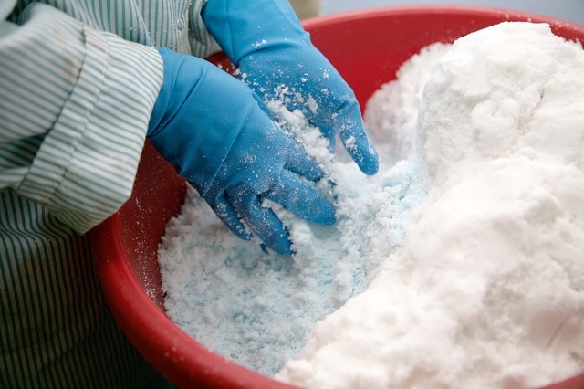
Figure 1. Zeolite. Image Credit: Fotosr52/shutterstock.com
X-ray diffraction is a powerful tool for determining the structure of crystalline materials – this is most often used for identification of unknown samples, such as in geology and earth sciences, or for studying protein structures in life sciences. It can also be used to determine the unit cell dimensions of a known crystal, or to measure the purity of a sample.
The technique is typically carried out on a single crystal, but because not all substances easily form good quality crystals, powder X-ray diffraction (PXD) is sometimes used instead.
Powder X-ray diffraction is particularly useful for substances like proteins, which are hard to crystallise, and zeolites, which are only available as powders. It can also be used for superconductors, which readily form powders, and protein drug complexes which easily form polycrystalline powders made of many small crystals in different phases. Because of this flexibility, PXD is becoming increasingly popular, despite the fact it offers less detailed information than single crystals, and it is harder to use to accurately determine bond lengths and angles.
Challenges of Structure Determination with PXD
Crystalline domains are randomly oriented in powder samples, which makes them particularly hard to study using X-ray diffraction. The main challenge PXD presents is that diffraction spots from powders are compressed from three dimensions in the reciprocal space to one dimension in the diffraction patterns. This means the data collected contains overlapping peaks, so the intensities of individual reflections are not known.
This makes the process of indexing to determine the correct crystal lattice constants particularly difficult, especially for challenging samples – for example, crystals with high-order symmetry or large unit cells, or macromolecules such as proteins, which are composed mainly of light atoms and do not exhibit very strong diffraction.
Overcoming this hurdle is fundamental to successfully using PXD for structure determination. One strategy is to develop more advanced X-ray optics, which increases the resolution of the diffraction data. Instrument manufacturers are working towards providing higher and higher resolution optics in laboratory instruments – however, the very best quality diffraction data is obtained at synchrotron facilities.
These are large particle accelerators which produce x-ray beams of extremely high intensity and quality, sufficient to determine the structures of even small protein molecules by powder diffraction.
Because these solutions are often impractical, at least for routine analysis, the PXD technique is more often used for simpler crystal structures, or to get coarse features of complex ones.
Solving Structures Ab Initio
The problem of overlapping peaks makes it particularly hard to solve structures ab initio (from first principles), because this relies upon being able to assign intensities to individual reflections. In recent years, the reduced cost of running advanced indexing and phasing algorithms has made ab initio PXD structure determination much more widely available.
Once indexing is complete, and the symmetry of the unit cell is known, the amplitude and phase data in reciprocal space must be converted into an electron density map that can be interpreted to give the full chemical structure.
As the phase cannot be measured directly, most approaches involve making an initial estimate, then refining the density map until it matches the input data as closely as possible. This can be done using a variety of algorithms, which can carry out calculations in reciprocal or real space. For example, ITO uses either direct or Patterson methods to solve the phase problem. DOVCOL generates random simulated structures in real space, and uses database searches and simple geometric modelling to find likely structures by minimizing energy.
Charge flipping is a relatively new method which is quite simple mathematically – as the name suggests, it reverses the sign (+/-) of any pixel in the initial charge density map below a threshold, then performs a series of transforms through reciprocal space. This iterative loop is repeated until a structure is identified. Because of its simplicity, this algorithm can identify even quite complex structures relatively quickly.
Other more sophisticated methods operating in real space (or direct space) can also be used to calculate electron density maps. These include Monte Carlo, which uses a large number of randomly generated input parameters to iteratively optimize a model, and genetic algorithms, which similarly start with a set of random structures, but mimic the biological processes of mutation and reproduction to find the final structure through an analog of natural selection.
Once there is enough information from one or more of these methods to make a reasonably confident estimate of the initial structure, a process of optimization follows, to tweak the model and produce an accurate structure for the crystal. This is most often carried out using the Rietveld method, which is a least-squares approach to fine-tune the model so that it matches the observed data as closely as possible.

Figure 2. Chemical production. Image Credit: Anna Jurkovska/shutterstock.com
By combining this approach with data from complimentary analysis techniques such as NMR spectroscopy, many unknown crystal structures can now be determined from PXD data. This is a great asset in many fields where high-quality crystals are not always available – for example, in the pharmaceutical industry where it is necessary to target specific properties when designing new molecules.
PXD is also proving useful for studying novel solid state chemistry and soft materials. Metal-organic frameworks, a new class of porous material of interest for catalysis, adsorption, and chemical sensing, have been studied extensively using this technique. The ability of powder X-ray diffraction to identify and characterize a wide range of substances gives it an important place in materials science research.
About Pittcon
 Pittcon, a leading conference and exposition for laboratory science, will take place between the 6th and 10th of March in Atlanta, Georgia. With an impressive technical program, the event will feature more than 2000 presentations. Among these, topics will include X-ray diffraction as a powerful tool for determining the structure of crystalline materials.
Among the 841 exhibitors at Pittcon will be Thermo Scientific, world-leaders in the field of X-ray diffraction. Thermo Scientific offer a wide range of systems covering a full spectrum of X-ray diffraction techniques.
Visit pittcon.org for more information.
References and Further Reading