Advanced Raman filters can be employed in Raman spectroscopy to deliver immediate data on biological tissue. These filters have great potential in research, medicine, and biomedical engineering and can be adapted to suit various applications.
Basic Raman spectroscopy setups can only provide basic information. More in-depth analysis is required in many cases, such as biomedical and bioanalytical applications.
Advanced Raman spectroscopy utilizes precise configurations and carefully selected filters to maximize the information the technique provides. Different biological samples require different laser wavelengths and filters. Therefore, it is important to determine the correct parameters for specific applications.
Raman Spectroscopy
In Raman spectroscopy, a sample is typically illuminated by a laser beam, and the scattered radiation is recorded and analyzed. The light scattering observed is classified as either Rayleigh scattering (at the laser wavelength) or inelastic scattering.
Inelastic scattering provides information on the sample, whereas Rayleigh scattering has a much higher intensity. A laser blocking filter (a notch, edge pass or band pass filter) separates the Rayleigh scattering from inelastically scattered light, resulting in the Ramen signal.
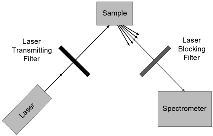
Image Credit: Avantier Inc.
A Raman scattering signal can reveal a sample's molecular makeup and even provide information on the intermolecular bonds.
In the past, holographic gratings and multiple dispersion stages separated out the Raman scattering, which was then collected by photomultipliers. Notch or edge filters are preferred and provide a much clearer signal.
Real-Time Tissue Analysis in Neurosurgery
Raman spectroscopy is ideal for real-time tissue analysis during neurosurgery. The technique provides immediate data, is non-destructive, and can be used to view tissue at the surface as well as provide a chemical fingerprint of cells several layers deep.
Residual tumors are a small number of cancerous cells that remain after removing the bulk of a tumor. They are one of the main reasons for neurosurgery failures. Therefore, it is paramount that healthy and cancerous tissue is differentiated during surgery.
Raman spectroscopy provides a means to distinguish between healthy tissue, pathological tumors, and necrosis. Raman techniques such as CRS, CARS and SRS microscopy can be implemented to provide a stronger signal and rapid imaging.
Osteoporosis Diagnosis
Osteoporosis leads to reduced bone quality, bone fragility, and low BMD, but historically, no medical diagnostic method has been capable of assessing the bone quality of a living subject.
Raman spectroscopy has the potential to provide such a diagnosis. Testing on mice has demonstrated that Raman spectroscopy can provide real-time data on the evolution of the carbonate/phosphate and mineral/matrix ratios that occur during the onset of osteoporosis.
Such techniques are expected to improve our understanding of osteoporosis's effect on human health and aid efforts to reverse or stop the bone deterioration involved.
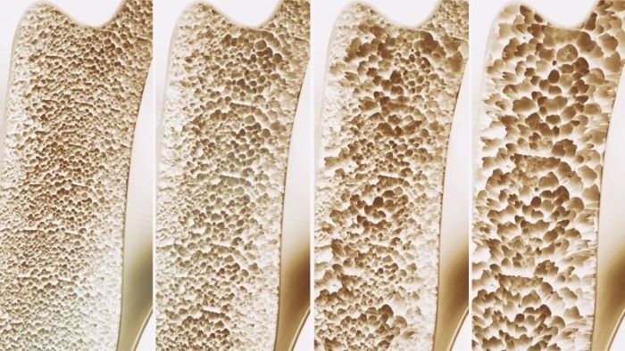
Raman spectroscopy can be used to gauge bone density and bone quality, critical markers of osteoporosis. Image Credit: Avantier Inc.
Optimal Optical Filters for Specific Applications
Avantier is a company that produces custom Raman edge, Bandpass, and notch filters optimized for blocking the laser line and transmitting anti-stokes Raman scattering.
The company also supplies laser line filters that transmit the exact wavelength of laser light required to illuminate a sample while blocking other radiation that could potentially degrade the signal-to-noise ratio.
Avantier engineers and designers are available for consultation to help clients determine the ideal wavelength ranges, optical density, and type of filter required for any given application. Assistance can also be provided to determine the ideal configuration and angle of incidence for specific systems.

This information has been sourced, reviewed, and adapted from materials provided by Avantier Inc.
For more information on this source, please visit Avantier Inc.