Ureteroscopes are flexible medical devices designed to facilitate diagnosis and treatment by enabling access to the kidney and urinary system.
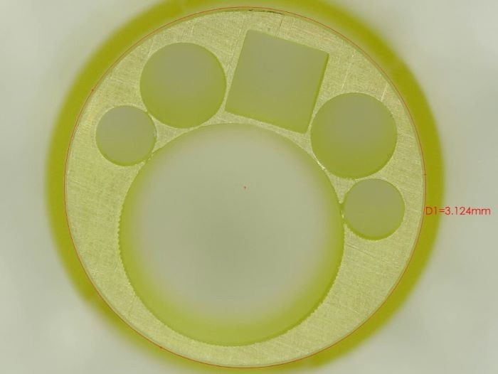
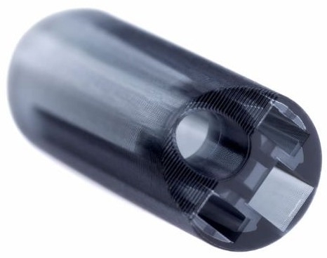
The endcap is central to this innovation. This component features a seamlessly integrated camera sensor, fiber-optic illumination, and a series of working channels designed for tools and irrigation.
3D printing is highly effective for producing ureteroscope endcaps, given the device's compact design with a diameter of just 3 mm. These components require extremely thin walls, approximately 50–75 µm thick, extending along the 5 mm length of the part.
Conventional manufacturing methods fall short or prove too costly when it comes to producing the miniature features required for these components. Consequently, micro 3D printing emerged as the only practical solution for efficiently and cost-effectively creating end-use parts with the necessary tolerances and resolution for this application.

Image Credit: Boston Micro Fabrication (BMF)
Ureteroscope with 3D printed end cap - unachievable using molding due to precision required
Video Credit: Boston Micro Fabrication (BMF)

Image Credit: Boston Micro Fabrication (BMF)
Using Micro 3D Printing to Develop a Single-Use Scope for Endourology
RNDR Medical has developed an innovative single-use scope for endourology designed for direct visualization and navigation within the urinary tract. This device supports the diagnosis and treatment of conditions such as kidney stones, urothelial carcinoma, and pyeloscopy procedures, providing therapeutic access to the renal pelvis and kidneys.
The ureteroscope is equipped with a digital high-definition camera and illumination system for clear visualization, alongside fluid irrigation, to maintain image clarity. Additionally, a working channel allows for the passage of therapeutic tools, such as lithotripsy fibers and retrieval baskets, to facilitate kidney stone treatment.
A critical component of the device is the distal tip, which houses the camera chip and illumination source, manages fluid paths for irrigation, and connects the working channel to the external anatomical environment.
This tip must meet stringent precision requirements, ensuring all components are securely housed and sealed to prevent fluid ingress, all within a compact diameter of just 0.130 inches. The tip’s exterior geometry is also designed to be atraumatic, minimizing potential harm as it advances through the anatomy.
Accommodating these design elements within such a small and precise profile requires a component with intricate 3D geometry, tight tolerances, and thin walls—specifications traditionally achievable only through specialized micro-molding techniques.
However, with annual production volumes in the tens of thousands, the high cost of micro-molding would result in a slower return on investment, presenting a challenge for scalable manufacturing.
Acknowledgments
Produced from materials originally authored by Boston Micro Fabrication.

This information has been sourced, reviewed and adapted from materials provided by Boston Micro Fabrication (BMF).
For more information on this source, please visit Boston Micro Fabrication (BMF).