Protein crystals have many uses ranging from basic research of the proteins themselves to controlling drug delivery, advanced drug design and even in bioseparation processes.

Most protein crystals are grown using a vapor diffusion process, such as the hanging or sitting drop methods. However, salt crystals are often commonly found alongside the protein crystals. Using normal imaging techniques, it is difficult to differentiate the two types of crystals. However, protein readily absorbs light at 280 nm and fluorescence in the near UV. While imaging the intrinsic protein fluorescence can differentiate the protein from the salt crystals, the technique is slow, subject to contamination effects and is limited in the information it can provide.
Even the well plates in which the crystals are grown may fluoresce. Ultraviolet microspectroscopy, on the other hand, is fast, highly accurate and provides much more information about the crystal other than just the fact that it is made up of protein.
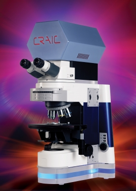
UV microspectrophotometers, such as the QDI 2010™ from CRAIC Technologies, Inc., are able to rapidly differentiate protein from salt crystals by both absorbance and fluorescence spectroscopy. More importantly, they can also take the next step to qualify the crystal once it has been located and identified. The procedure is simple: a spectrum is acquired of a microscopic crystal. If it is salt, it will not absorb light at 280 nm. However, the protein does exhibit a strong absorbance peak near that wavelength.
From the spectra, the concentration of the protein in the crystal can be determined or even if the crystal is contaminated. Due to the inherent flexibility of microspectrophotometers, the crystals may be both imaged and microspectra™ acquired of crystals as small as a micron. Able to operate in the visible and NIR regions, in addition to the ultraviolet, these systems can do much more than just locating crystals. The ability to use microspectra™ to rigorously qualify the crystal can save valuable time by only selecting viable crystals for the next step of a procedure.
For more information on drug delivery, click here.