The worldwide Raman Imaging Community met from 26. – 27. September at the 9th Confocal Raman Imaging Symposium in Ulm, Germany to discuss the latest developments in the field. Featured at this year’s event were the application possibilities of high-resolution Raman microscopy in pharmaceutical research as well as materials, geo-, and life sciences.
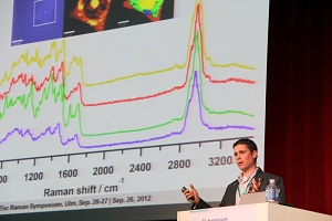
 Dr. Diedrich Schmidt from the North Carolina A&T State University, USA, presenting results of single cell measurements.
The keynote lecture by Prof. Sebastian Schlücker from the University of Osnabrück, Germany, on the basics of Raman spectroscopy and its application in microscopy provided a broad interactive look at the classical and quantum-mechanical description of the Raman effect. The specific requirements for highly sensitive and diffraction-limited Raman imaging in terms of an optimized microscope setup were highlighted by Dr. Olaf Hollricher, WITec Managing Director R&D. Dr. Thomas Dieing, WITec Director Customer Support, underlined in his presentation on the combination of Raman with structural surface imaging, the necessity of a modular instrument platform in order to perform AFM and TrueSurface Microcopy with a single instrument.
In the first application-related talk, Dr. Thomas Beechem from Sandia National Laboratories discussed the analysis of stress and material interactions between the individual layers of a bi-layer graphene sheet. Dr. Juan Romero from CSIC, Madrid, Spain, talked about the importance of ceramic materials in everyday life as well as high technology while showing various examples of how Raman imaging is beneficial for the characterization of ceramic materials. The advantages of 3D analysis were illustrated Dr. Barbara Cavalazzi, University of Johannesburg, reporting on the investigation of fossilized traces of life on earth. The importance of Raman imaging in Life Science as a label-free method was discussed by Dr. Diedrich Schmidt from the North Carolina A&T State University, USA, presenting results of single cell measurements. Dr. Klaus Wormuth from the University of Cologne, Germany used Raman microscopy to achieve a better understanding of drug delivery systems. In the related presentation that followed, Dr. Maike Windbergs from the University of Saarbrücken, Germany, showed methods that can successfully characterize pharmaceutical systems with a special focus on results obtained with TrueSurface Microscopy.
The poster session was commenced by Dr. Dieter Fischer, from the Leibnitz Institute for Polymer Research, Dresden, Germany with his presentation on temperature-dependent Raman imaging of polymer blends, which was selected as a plenary presentation among the abstracts of the contributed poster session. Nearly 20 contributions for the poster session allowed intense discussion on the scientific results of the participants. The poster session was well-attended and the contributions were favorably received.
During the dinner talk Prof. Fernando Rull from the University of Valladolid, Spain, reported on the Raman spectrometer which will be part of the European ExoMars 2018 mission, pointing out that the instrument design for space applications requires additional evaluative research to be successful.
The main focus of the second symposium day was the operation of the instruments. In the WITec application lab, the attendees were able to see live demos of 3D Raman imaging experiments combined with AFM or topographic Raman Imaging on large samples. The necessary data evaluation software was presented as well.
In summary the participants of the symposium were presented with a varied and detailed view of the current status of Raman imaging knowledge. The high scientific level of the presentations was well-regarded as reflected in the 100 % of attendees who rated the symposium as “Very Good” in a final anonymous feedback poll.