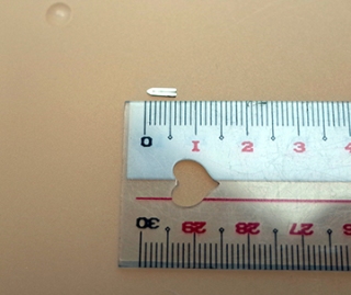
Researchers at the Kobe University have developed a safe, dissolvable surgical clip made of a magnesium alloy. This unique clip has the potential to reduce risk of complications after surgeries, and difficulties faced while performing diagnostic imaging.
 The surgical clip
The Division of Hepato-Biliary-Pancreatic Surgery at the Kobe University Graduate School of Medicine had collaborated with the Division of Mechanics and Physics of Materials at the Kobe University Graduate School of Engineering for developing this clip. Currently, titanium is the most widely used material in most surgical clips. A single surgical procedure may require as many as 30 to 40 clips. However, these clips remain within the patient’s body even after the wound healing is complete, and it may lead to complications. Further, when imaging using MRI (magnetic resonance imaging) or CT (computed tomography) is performed around the wounds, these clips reduce the quality of the images obtained.
These innovative clips are made of a magnesium alloy, which includes zinc and calcium that contribute to a better microstructure, formability and fastening ability. Further, these clips are just 5mm in size. All of these qualities are desirous of clips needed for surgical procedures.
As part of this study, in vivo studies were conducted to evaluate the clip’s safety and functionality. The research team performed an implantation study in a subcutaneous mouse model to evaluate the safety of the clip. They observed that the dissolution of the clip resulted in the generation of a small amount of gas and inflammation did not occur in the surrounding tissues after a period of 1 to 12 weeks.
This would probably mean that the clip might not be causing significant adverse effects. After a period of 12 weeks, tests revealed that the levels of magnesium in the blood remained within the normal range. Further, the clip had dissolved by nearly 50% and would probably dissolve and come out of the patient’s body within a year.
The team evaluated the clip’s functionality in a rat model. Here, they removed a partial liver and occluded the biliary duct, hepatic vein, hepatic artery, and portal vein with the clip. They monitored the rat for a period of 8 weeks, and they observed that the rat did not develop any problems. This could mean that the clip was functioning in the correct manner. The team performed micro-CT scanning on the rat and the mouse, and found that the organs could be observed as there was no degradation in the image quality.
We will conduct further in vivo studies and a clinical study within 2 to 3 years. Kobe University works toward the development of new medical devices. We will continue to promote collaboration between the Graduate Schools of Medicine and Engineering.
Professor MUKAI Toshiji, Department of Mechanical Engineering, Graduate School of Engineering
References