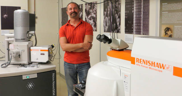
The University of California, Los Angeles (UCLA), USA, combines Raman microscopy with scanning electron microscopy (SEM) to study archaeological textiles and fibres.
UCLA use a Renishaw SEM-SCA system to study textiles and fibres, non-destructively. The full paper describing the work has been published by the Journal of Raman Spectroscopy (John Wiley & Sons, Ltd.).

The Department of Materials Science and Engineering at UCLA delivers novel and highly innovative research that advances basic and applied knowledge in materials. This is achieved through interactions with the external community, and educational outreach and industrial collaborations.
This is illustrated by a paper joint-authored by scientists from UCLA, the Los Angeles County Museum of Art, the Cotsen Institute of Archaeology and instrumentation manufacturer, Renishaw1.
The paper “New Advancements in SERS Dye Detection Using Interfaced SEM and Raman Spectromicroscopy (µRS)” is published in the Journal of Raman Spectroscopy. It describes how a SEM may be interfaced with µRS to provide a means of non-destructively identifying organic compounds in complex samples, such as single fibres, by extractionless analysis. This enables the characterisation of dyes from reference collections and archaeological textiles.
The lead author of the work is UCLA scientist, Dr Sergey V Prikhodko. He describes why Raman Spectromicroscopy has become a powerful analytical technique for the study of artistic, historical and archaeological materials:
“As a non-destructive method that may also be interfaced with other techniques, it makes the sample reusable for subsequent analyses after performing µRS. In situ morphological characterization, elemental identification and structural analysis integrates µRS with SEM and energy dispersive X-ray spectroscopy (EDS) in the ‘hyphenated’ SEM-EDS-µRS system, at UCLA. This vital capability was brought together with the Renishaw SEM-SCA interface, where a Nova 230 SEM (FEI) is coupled to the Renishaw inVia confocal Raman microscope to provide structural and chemical analysis in situ.”
Dr Prikhodko’s results on samples of various fibres clearly illustrate the potential of this non-destructive method. Interfacing SEM with µRS provides a very powerful tool to analyse unique and irreplaceable samples and artefacts quickly and in a cost-effective manner.
Please visit www.renishaw.com/SEMRaman for further details of Renishaw’s inVia confocal Raman microscope and the SEM-SCA interface.
Reference
1. Prikhodko, S. V., Rambaldi, D. C., King, A., Burr, E., Muros, V., and Kakoulli, I. (2015), New advancements in SERS dye detection using interfaced SEM and Raman spectromicroscopy (μRS). J. Raman Spectrosc., doi: 10.1002/jrs.4710.
About Renishaw
Renishaw is a world leading engineering technologies company, supplying products used for applications as diverse as jet engine and wind turbine manufacture, through to dentistry and brain surgery. It employs over 4,000 people globally, some 2,600 of which are located at its 15 sites in the UK, plus over 1,400 staff located in the 32 countries where it has wholly owned subsidiary operations.
For the financial year ended June 2014 Renishaw recorded sales of £355.5 million of which 93% was due to exports, the largest markets being the USA, China, Germany and Japan.
The Company's success has been recognised with numerous international awards, including eighteen Queen's Awards recognising achievements in technology, export and innovation. Renishaw received a Queen’s Award for Enterprise 2014, in the Innovations category, for the continuous development of the inVia confocal Raman microscope. For more information visit www.renishaw.com