Mar 23 2017
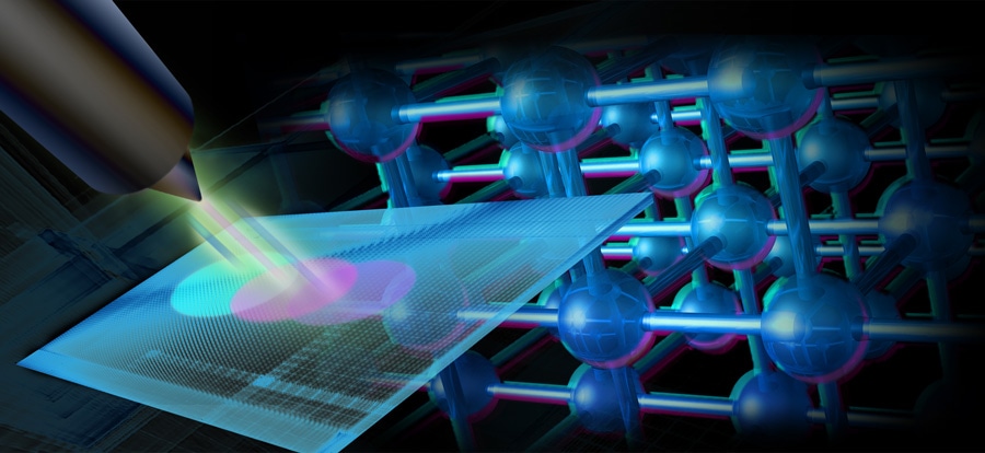
 Scientists have used a new X-ray diffraction technique called Bragg single-angle ptychography to get a clear picture of how planes of atoms shift and squeeze under stress. (Image by Robert Horn/Argonne National Laboratory.)
Under stress, everything reacts differently - even the relatively orderly atoms in crystals. If scientists could get a clear picture of how planes of atoms move and squeeze under stress, they will be able to use those properties in order to provide promising technologies, such as next-generation semiconductor components and nanoelectronics, with additional functionalities or speed.
However, this picture requires new techniques for imaging atoms in materials and their behavior in varied environments.
A new form of imaging that employs X-ray diffraction patterns, called single-angle Bragg ptychography, has been developed by scientists in a recent collaborative study from the Institut Fresnel, IBM and the U.S. Department of Energy’s (DOE) Argonne National Laboratory.
Despite the fact that Bragg ptychography and particularly X-ray diffraction have been around for quite some time, single-angle Bragg ptychography permits for much easier reconstruction of 3D data about how a material is affected by strain.
In X-ray diffraction, incoming X-rays are scattered by the atoms within a material, developing a signal on a detector. Identifying the contribution of a specific small region of the lattice to the overall signal could be difficult as a number of overlapping diffraction events take place simultaneously.
In order to compensate for this, a method called Fourier analysis was used by scientists. This method transforms the overall signal to a series of waves with valleys and peaks that correspond to the relative intensities of different parts of the signal.
In order to really see and understand the strain in real space, you need information on both intensity and the phase. What we needed was a trick to retrieve the missing phases of the diffraction pattern.
Stephan Hruszkewycz, Material Scientists, Argonne National Laboratory
It is possible to understand phase by imagining waves lapping onto the shore after a handful of rocks is thrown into a still pond. It is also possible to “watch the wave backwards” by reconstructing the sizes and positions of all the rocks when they hit the water.
This is brought about by measuring the height of waves at the shore and also their time of arrival. However, X-ray detectors only measure the height of the waves; phases, i.e. when the wave reaches the shore must be recovered through other means.
The trick used by the authors comes from ptychography, which is a technique that uses redundant sampling from the same region of the crystal in order to recover phase information. The team succeeded in extracting information about the phase by shifting the X-ray beam only slightly, followed by imaging as much as 60% of the same real space existing between beam positions.
“In essence, by having a lot of the same information encoded in neighboring samples, it constrains the possible configurations of the crystal in real space,” Hruszkewycz said.
However, the real advance came from the positioning of the beam itself and not from information gathered through diffraction. The researchers were able to extract extra information about how the material was affected by the strain in three dimensions as they knew exactly where the beam was placed and the angle at which the atomic planes of the crystal would scatter the X-rays.
Most diffraction techniques, including some ptychographic ones, really only give a 2D representation of the sample of interest. This technique also makes fewer requirements in terms of the instrument technology than comparable techniques for generating 3D information about materials.
Stephan Hruszkewycz, Material Scientists, Argonne National Laboratory
An article based on the study, “High-resolution three-dimensional structural microscopy by single-angle Bragg ptychography,” was published in November in the online edition of Nature Materials.
The U.S. Department of Energy’s Office of Science (Office of Basic Energy Sciences) funded the work. The Hard X-Ray Nanoprobe Beamline, operated by Argonne’s Center for Nanoscale Materials at Argonne’s Advanced Photon Source, was used by the researchers. Both the Center for Nanoscale Materials and Advanced Photon Source are DOE Office of Science User Facilities.
Martin Holt and Paul Fuoss, Argonne scientists, also contributed to the study, along with researchers from IBM and France.