From CSEMReviewed by Mychealla RiceJun 28 2022
Complex 3D cell models are revolutionizing personalized medicine in drug testing and tissue engineering. Implementing these mini-organs in the applied research and industrial landscape requires new tools for process standardization and parallelization. HistoBrick is a new tool that paves the way toward high-throughput and standardized micro-histology.
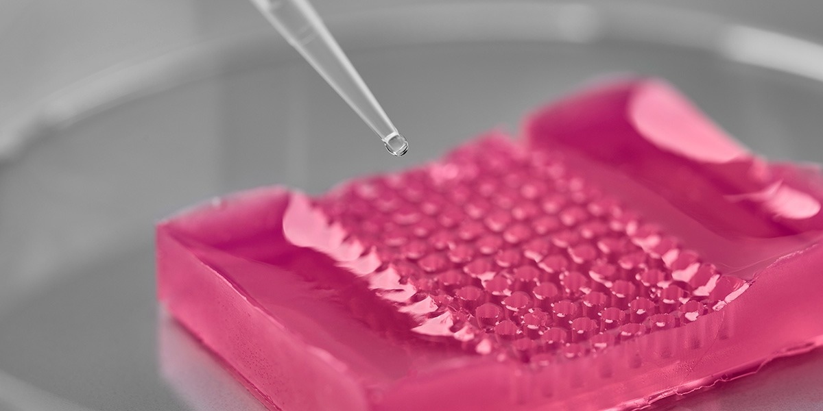
 HistoBrick: a textured hydrogel-based plate enabling sectioning of 96 micro-tissue arrays throughout the paraffin-embedding histology process. Image Credit: CSEM
CSEM Tools for Life Sciences is developing solutions for all life stages of micro-tissues, organoids, and tumoroids: production, sorting, positioning, maturation, monitoring, and analysis. The examination of the anatomy of the cells produced in 3D cell models is of particular importance throughout the entire value chain. A new discipline, so-called micro-histology, is addressing this current need of aligning hundreds of organoids in one focal plane for imaging and analysis.
HistoBrick, a new micro-histology tool
In collaboration with the School of Life Sciences FHNW (Prof. Laura Suter-Dick), with its advanced expertise in cell biology and in vitro toxicology, CSEM Tools for Life Sciences has developed a new approach to align organoids. HistoBrick offers precise co-planar positioning of 96 micro-tissues before dehydration, paraffin embedding, and sectioning. All micro-tissues are sectioned simultaneously at the same level resulting in reduced processing and analysis times at high yield.
Official publication in Nature Scientific Reports
Sarah Heub, Fatemeh Navaee, Daniel Migliozzi, Diane Ledroit, Stéphanie Boder-Pasche, Jonas Goldowsky, Emilie Vuille-Dit-Bille, Joëlle Hofer, Carine Gaiser, Vincent Revol, Laura Suter-Dick, and Gilles Weder, “Coplanar embedding of multiple 3D cell models in hydrogel towards high-throughput micro-histology”, Sci Rep 12, 9991 (2022). Nature Scientific Reports (doi:10.1038/s41598-022-13987-4)