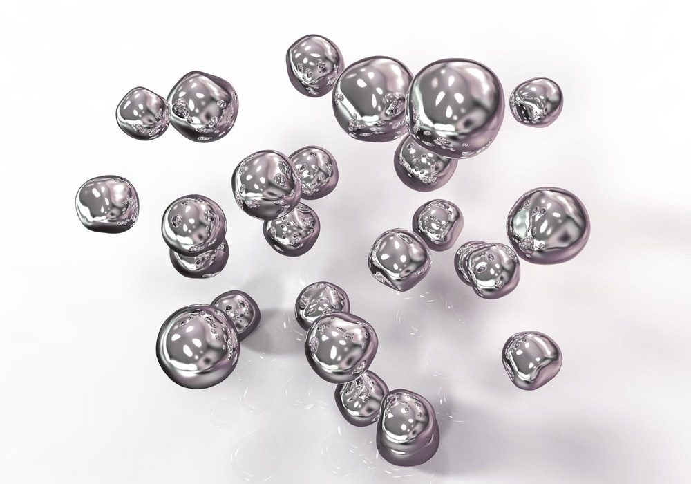
A recent article published in Scientific Reports proposed the fabrication of a silver nanoparticle (NPs)-based poly(vinyl alcohol) (PVA) and polycaprolactone (PCL) membrane. It exhibited antibacterial properties against gram-negative Escherichia coli (G-E. coli) and gram-positive Staphylococcus aureus (G+S. aureus) and no cytotoxicity on the Vero cell line.

Image Credit:Kateryna Kon/Shutterstock.com
Background
Chronic non-healing wounds pose a serious risk of infection and subsequent health complications. Recently developed multilayered wound dressings, consisting of antibacterial agents and fibrous matrices, provide a hygroscopic adhesive layer for new cell generation. Silver dressings are also used when an infection is established or when healing is delayed due to critical colonization or pre-infection.
Nanofiber mats prepared by electrospinning also find applications in wound dressing. Materials used for electrospinning and electro-spraying in the medical field include synthetic polymers such as water-soluble PVA and water-insoluble PCL. These materials are used as carriers for bioactive agents like NPs or as templates or artificial fibers to form fibrous products.
The electro-spun three-dimensional fiber structures fabricated using these biocompatible polymers closely mimic the natural extracellular matrix, essential for optimal cell attachment and growth. Thus, the researchers prepared a polymer-metal-based membrane using electrostatic processes for wound dressing and other medical applications.
Methods
In this study, maleic acid and silver nitrate were used to synthesize Ag NPs with a size of 8 ± 3 nm. The morphology and size of these Ag NPs were characterized using a high-resolution transmission electron microscope (HRTEM), and the resulting data was analyzed using fast Fourier transformation (FFT) in ImageJ software.
The synthesized Ag NPs were anchored to an electrospun PCL matrix via electro-spraying PVA. These PVA+Ag materials were prepared by 15, 30, 45, and 60 minutes of spraying to obtain gradually increased Ag NP content in the PVA droplets.
Before the electro-spraying process, a 10 wt.% solution of solid PVA was diluted in demineralized water, while a 10 wt.% solution of solid PVA was diluted in the prepared Ag colloid (PVA+Ag).
The PVA droplets and PCL fibrous samples were investigated by scanning-transmission electron microscopy (STEM) equipped with energy-dispersive X-Ray spectroscopy (EDS). Subsequently, the electro-spun fiber size distribution was obtained by image analysis. For structural characterization using X-Ray diffraction (XRD), a thin layer of the colloidal sample was deposited on a glass plate.
EDS analysis confirmed the presence of Ag, while the release of Ag from fibrous samples was determined through a spectrometer.
The antibacterial activity of different PCL/PVA+Ag membranes was evaluated against G+S. aureus and G-E. coli bacterial cultures obtained from the Czech collection of microorganisms.
Finally, aware of the possible cytotoxicity of Ag NPs, the researchers examined their effect on the Vero cell line. They then assessed morphology, vacuolization, detachment, cell lysis, and membrane integrity under an optical microscope using qualitative cytotoxicity evaluation.
Results and Discussion
As revealed by HRTEM analysis, Ag NPs were primarily spherical and oval-polygonal. When attached to the PVA matrix, these formed droplets, which served as an Ag NP carrier. The number of attached PVA+Ag droplets also increased with longer electro-spraying durations, reaching the highest accumulation for the PCL/PVA+Ag_60 sample (sprayed for 60 minutes).
During antibacterial tests, as a water-soluble polymer, PVA dissolved with humidity, causing a burst of Ag NP release. Bacterial growth inhibition was observed after three hours, with a more rapid effect against G+S. aureus compared to G-E. coli. This can be attributed to the smaller size of S. aureus, allowing fast penetration of Ag NPs.
The PCL electro-spraying time significantly influenced the antibacterial properties of PCL/PVA+Ag spheres, as evidenced by the efficiency of the PCL/PVA+Ag_60 sample. It inhibited the growth of both bacterial species after three hours of incubation, reaching 100 % inhibition after six hours.
However, the control samples comprising pure PCL and PCL/PVA_60 without Ag NPs demonstrated no antibacterial effect against either bacterium.
Although Ag is an acknowledged antibacterial agent against both G+ and G- bacteria, some bacterial strains develop rapid resistance to them after repeated exposure. They can also exhibit cytotoxic effects on mammalian cells at certain concentrations. However, the cytotoxicity test results on the Vero cell line indicate non-toxicity of all samples prepared in this study.
Conclusion
This study connected the nanofiber and spherical polymer structures of PCL. Electro-spraying effectively delivered antibacterial agents during postprocessing to the PCL matrix. The resultant silver-based materials exhibited 100 % inhibition of G+S. aureus compared to G-E. coli within six hours while showing non-cytotoxic effects on the Vero cell line.
The proposed material preparation method allows for various modifications to enhance the antibacterial efficiency, such as the preparation of a multilayered structure with possible sustained release of antibacterial agents.
The morphology, size, and surface properties of active Ag NPs can also be altered, and their concentration can be increased by extending the spraying time of the PVA+Ag polymeric solution.
Combining antibacterial properties and non-cytotoxicity, such materials hold promise for various medical applications.
Journal Reference
Vilamová, Z., et al. (2024). Silver-loaded poly(vinyl alcohol)/polycaprolactone polymer scaffold as a biocompatible antibacterial system. Scientific Reports. doi.org/10.1038/s41598-024-61567-5
Disclaimer: The views expressed here are those of the author expressed in their private capacity and do not necessarily represent the views of AZoM.com Limited T/A AZoNetwork the owner and operator of this website. This disclaimer forms part of the Terms and conditions of use of this website.