Sponsored by HORIBAJun 14 2018
Raman spectroscopy can be combined with optical confocal microscopy to generate a new analytic technique called Raman microscopy. Light microscopy has several advantages: it can be used to observe living cells, and thus to watch many types of biological activity including feeding, cell division and locomotion.
In vivo stains can be added to observe how cells take up colored pigments. Raman microscopy cannot achieve these tasks successfully in real-time, both because image acquisition takes a long time, and because specimens for an electron microscope need to be fixed and dehydrated, typically resulting in cell death. In addition to the above benefits, optical microscopes have also been found to be of great use when studying various fields of science and medicine.
Advantages of Raman Spectroscopy
Some important advantages of Raman spectroscopy include:
The Ability to Carry out a Detailed Analysis of Chemical and Molecular Structure
Raman spectroscopy studies vibrational motion within individual chemical bonds. The Raman spectrum is thus rich in data which helps to determine the chemical structure of the analyte, its composition, and its identity if unknown, by matching its spectral profile with large Raman spectrum databases.
The Availability of Less Obvious Information
Raman spectroscopy not only identifies the analyte in a general way and reveals its characteristics, but brings out more hidden data such as such as the crystalline structure, polymorphous state and phases, in addition to inherent strain or stress on the molecular structure, folding of proteins and the presence of hydrogen bonds.
Speed
Raman spectroscopy operates within a few seconds to yield a high-quality spectrum. It does not need to use a prepared sample which allows high throughput.
No Need to Prepare the Sample
There is no need to subject the sample to grinding, dissolution, pressing or glass formation before Raman spectroscopy. This means that samples can be used as received, whether slurry or liquid, gas, or powder.
Non-Destructive Technique
Raman spectroscopy is a non-destructive method and does not even require contact with the specimen. The lack of any need for sample preparation means that samples which have historic importance, such as antique pigments or forensic samples, may be analyzed while still remaining intact and available for testing by other methods if required.
Good Spatial Resolution
Raman confocal microscopy combines a microscopic examination of high sensitivity with the ability to resolve details to below a few micrometers in size. This means that in practice, one but not another area of a sample can be analyzed, or single grains or particles of a specimen can be tested. Raman micro spectroscopy offers all these benefits as well.
Confocal Analysis
When a true confocal Raman microscope is used, there is spatial resolution covering a complete 3D range. This permits a single volume of a transparent sample to be analyzed. This property is extremely useful when it comes to analyzing samples which are in layers such as laminated polymers, inclusions like those frequently found in minerals or in glass, thin samples such as cells or tissues mounted on a substrate like a slide, and specimens contained in glass or plastic containers such as bottles of liquid.
Allows Analysis to be Performed In-Situ, In-Vitro and In-Vivo
Raman spectroscopy is both non-contact and non-destructive and therefore in-situ analysis can be performed on a specimen, such as analyzing a chemical reaction proceeding within its reaction vessel without disrupting the reaction in the slightest.
It can be carried out in the presence of water which makes it appropriate for in-vitro and in-vivo analysis of cosmetics applied on skin, the characterization of single cells in microbiologic tests, and examining interactions between a drug and different cell types during the process of designing drugs intelligently.
Raman Compared with Micro-FTIR
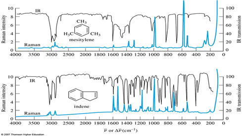
Raman microscopy often goes through comparisons with the gold standard spectroscopic technique for molecules, namely, FTIR. However, these techniques are better treated as complementing each other rather than as alternatives.

Raman FTIR Spectra from mesitylen (Image Credit: 2007 Thomson Higher Education)
Raman microscopy has some advantages over the use of IR technology, namely:
- Lack of interference due to the presence of solvents or cells, or because of prior sample preparation
- More specific, with narrow peaks
- Allows depolarization studies to be performed and even improves the results in some cases
- Can detect vibrational movement modes which are not IR-active
Raman Microscopy vs. Electron Microscopy
Raman microscopy is quite different from electron microscopy, because even though both are used to image physical and chemical structures on a microscopic scale in the study of biological and other materials, they are used in different ways and complement each other.
Raman microscopy has some advantages over electron microscopy, namely:
- Samples do not need to be prepared first
- The environment is simpler to handle because there is no need for a vacuum, for controlling the relative humidity, heat or cooling parameters
- Quicker analysis with higher throughput
- Rapid setup with less than ten minutes required to start analysis after switching on the system
- Ability to measure samples in multiple layers
- A much more compact workbench
Raman Spectroscopy and XRD
Raman spectroscopy is complementary to XRD when analyzing the crystal structure of a material.
Raman spectroscopy is superior to XRD in some ways:
- It allows the measurement of both crystalline and amorphous substances
- It allows analysis of even single grains or particles
- It can be performed under simpler conditions which do not require vacuum production, or control of relative humidity, heating or cooling
- The workbench is much more compact

This information has been sourced, reviewed and adapted from materials provided by HORIBA.
For more information on this source, please visit HORIBA.