lsuaneye / Shutterstock
X-ray crystallography is a powerful non-destructive technique for determining the molecular structure of a crystal. X-ray crystallography uses the principles of X-ray diffraction to analyze the sample, but it is done in many different directions so that the 3D structure can be built up. It is a technique that has helped to deduce the 3D crystal structure of many materials, especially biological materials.
When you think of X-ray diffraction (XRD), a 2D diffraction pattern comes to mind for most. The basic patterns generated in X-ray crystallography are still 2D diffraction patterns, but the key difference is that the sample is scanned in multiple directions. These diffraction patterns are then pieced together and refined multiple times so that they analyze and determine the molecular structure of the sample. It is even possible to do this for very large and complex molecules, with proteins being a key example.
Key Principles and Workings
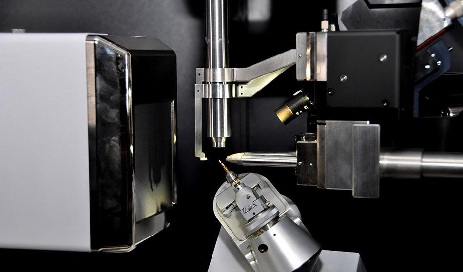
In the instrument, the sample is mounted on to a goniometer, which is used to position the crystal into specific orientations so that it can be analyzed from multiple angles. In some cases where the sample is impure and the crystal structure is not clear, the crystalline sample will need to be purified before analysis.
X-rays are generated from an X-ray tube, and they are then filtered so that they are monochromatic, i.e. of a single wavelength frequency. The atoms in the crystal refract the X-rays and the X-rays are elastically scattered on to a detector. Because they are elastically scattered, they have the same energy as the incident X-rays that are fired at the sample. This generates a 2D diffraction pattern of the crystal in a single orientation.
If the diffraction pattern is not clear, then the sample may not be pure and will be purified at this point. But other factors can prevent a diffraction pattern from being generated including a too-small sample (needs to be at 0.1 nm in each dimension), an irregular crystal structure, and the presence of any internal imperfections—such as cracks—in the crystal.
If the crystal is deemed to be ok, then the analysis and X-ray bombardment towards the sample continues. The sample rotates on the goniometer so that a series of 2D diffraction patterns are generated from various sides of the sample. The intensity is recorded at every orientation and the result is thousands of 2D diffraction patterns that correspond to different parts of the 3D structure. From here, a computational approach analyses the different diffraction phases, angles and intensities to generate an electron density map of the sample. The electron density map is used to generate an atomic model of the sample. The model is constantly refined to ensure that it is accurate, and once the final atomic model has been established, the data goes into a central database to act as a known reference.
Applications
In terms of applications, X-ray crystallography is used in many scientific fields. When it was first established as a useful technique, it was primarily used in fundamental science applications for determining the size of atoms, the lengths and different types of chemical bonds, the atomic arrangement of materials, the difference between materials at the atomic level, and for determining the crystalline integrity, grain orientation, grain size, film thickness and interface roughness of alloys and minerals.
Science has come a long way since then and while these areas are still important for analyzing new materials, it is now often used to identify the structure of various biological materials, vitamins, pharmaceutical drugs, thin-film materials and multi-layered materials. It has become one of the standard ways of analyzing a material if the structure is unknown across the geological, environmental, chemical, material science and pharmaceutical sectors (plus many others) due to its non-destructive nature and its high accuracy and precision.
Nowadays, it is often used to probe specific ways in how the structure of a material, drug, or substance will interact in certain environments. This is has become particularly useful across the proteomics and pharmaceutical sectors. Some of the specific areas that can now be probed with X-ray crystallography include measuring the thickness of films, identifying specific crystal phases and orientations that can help to determine the catalytic activity of materials, determining the purity of a sample, determining how a drug might interact with specific proteins and how the drug can be improved, analyzing how proteins interact with other proteins, for investigating microstructures, and for analyzing what amino acids are present in a protein which can help with determining how catalytically active an enzyme is. These are just a few specific examples as the use of X-ray crystallography is widespread.
Sources and Further Reading
Disclaimer: The views expressed here are those of the author expressed in their private capacity and do not necessarily represent the views of AZoM.com Limited T/A AZoNetwork the owner and operator of this website. This disclaimer forms part of the Terms and conditions of use of this website.