Marine pollution has become an ever-growing problem in today’s world. Microplastics have become the dominant form of plastic debris found in oceans globally and are found throughout most seawater samples.
Microplastics are specified as fragments of plastic measuring less than 5 millimeters in diameter, while nanoplastics are tinier still with diameters typically less than 0.001 millimeters. Penetrating the natural ecosystem in several ways including via wastewater and industry these plastics are found in a range of sources including clothing, cosmetics, cleaning products, and industrial processes.
Their minute size makes it possible for marine organisms to easily ingest them which allows the micro and nanoplastics to enter the food chain. Furthermore, certain additives used to treat the material, such as flame retardants and Bisphenol A, can lead to it being all the more toxic.
Recently, a discovery led by a group of researchers based at Arizona State University detected micro and nanoplastics in human tissues and organs (https://www.news-medical.net/news/20200818/Nano-and-microplastics-found-in-all-human-organs-and-tissues.aspx)

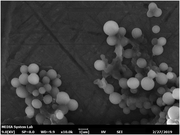
Figure 1. Microplastic particles separated from sea water and imaged on a Coxem EM-30AXPlus tabletop SEM. Image Courtesy of MEDIA System Lab (www.m-s.it)
Conventional methods of analysis usually isolate and study individual particles, which can be a laborious and time-consuming process. Therefore, as concern and interest in the identification and analysis of these pollutants increases, a fast and precise method to do so is needed. By using SEM to examine distribution and particle size and EDS to identify chemistry, it is possible to accelerate the analysis process, improve understanding of the severity of the problem, and create new techniques to help alleviate the problem in the future.
The Coxem EM-30 is an optimal tool for this kind of research and study. With a precision stage, the capacity for automated imaging and analysis of numerous specimen stubs, and a small footprint, the EM-30 is an economically affordable compact SEM for environmental and research labs.
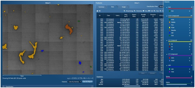
To demonstrate the capability of the machine, an experiment was conducted in which samples from the Tan Cheon River were taken. These samples were then vacuum filtered to eliminate all traces of water prior to being placed in the SEM. Subsequently, utilizing EDS to identify and analyze the individual particles with automated particle analysis, it was possible to analyze 435 fields of 2.75 mm2 each in just 2 hours and 40 minutes. Having discovered and detected 848 individual particles after initial analysis, each particle was then measured for chemistry and size (area and circumference).

Figure 2. Automated feature analysis is used to scan, identify, and classify microplastic particles.
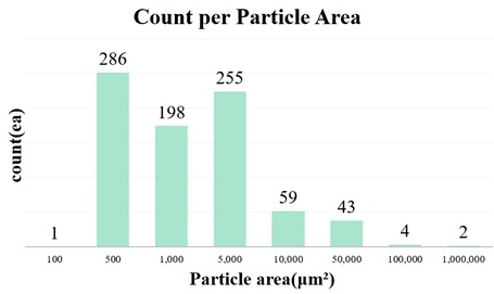
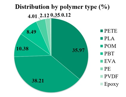
The data below illustrates that most particles discovered had surface areas somewhere between 500 to 5000 µm2, and the dominant type of plastics was Polyethylene terephthalate (PETE), frequently used in the production of plastic containers for liquids and foods as well as being a common material used in fibers for clothing. The other most-prevalent plastic found in the samples was Polylactic Acid (PLA), a popular bioplastic.

Figure 3. Particle size distribution binned by area.

Figure 4. Distribution by polymer type (identified by EDS).
Therefore, utilizing SEM in combination with EDS gives rise to an extremely effective tool when it comes to analyzing various pollutants, particularly microplastics. Making practical use of automated feature analysis can lead to the development of straightforward and accurate techniques to usher in rapid analysis for the classification of microplastic particles in water samples.

This information has been sourced, reviewed and adapted from materials provided by COXEM Co. Ltd.
For more information on this source, please visit COXEM Co. Ltd.