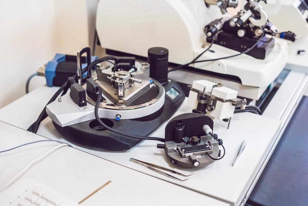
This article explores multi-frequency magnetic atomic force microscopy (MFM-AFM) and its utilization in detecting magnetic fields.

Image Credit: Elizaveta Galitckaia/Shutterstock.com
What is MFM-AFM?
A standard single frequency AFM is comprised of a boron-doped silicon (Si) or silicon nitride (Si3N4) cantilever with a length of a few micrometers and a single crystal diamond tip at the bottom of its free-end. It is used to measure linear interactions between tip and surface such as mapping the topography of surfaces with precision up to the nanometer scale.
The tip has a width of a few nanometers, which can oscillate vertically using a laser beam falling on the top of the cantilever and just above the diamond tip. Afterward, the laser beam reflects towards a photodiode. Any change in the position of the tip can be measured by the change in the path of the laser beam recorded by the photodiode. Other than a laser-photodiode-based scanning system, a system with piezoelectric material at the base of the cantilever and generated sinusoidal voltage can also be used to measure the deflection of the cantilever.
Dynamic-mode multi-frequency AFM (MF-AFM) is a step ahead of standard single-frequency AFM. It can simultaneously measure topography along with other modes of interactions between the tip and surface such as mechanical, electrical, magnetic, optical, and chemical. The frequency of the external signal (i.e. laser or sinusoidal voltage) can be varied to modulate with the oscillation of the cantilever.
It can be an amplitude modulation (AM) or a frequency modulation (FM). Based on amplitude or frequency modulation, the respective shift in the values of amplitude or frequency of the cantilever from their initial values to the resonance values can be used to measure both linear and non-linear interactions. The nonlinear interaction forces have a gradient along the path of oscillation of the tip.
Furthermore, MFM-AFM is specifically used for measuring localized magnetic fields on the surface of a substrate. Localized magnetic fields, also known as spin-orbit interactions, are formed by the interaction between the orbital motion of electrons around the nucleus of an atom and magnetic dipole moment due to the spinning of an unpaired electron.
More from AZoM: Analyzing Membrane Fouling with Atomic Force Microscopy
The tip of MFM-AFM consists of an additional ultra-thin layer of ferromagnetic material such as nickel (Ni). Usually, the Ni thin-film is deposited on a Si or Si3N4 wafer using the evaporation deposition method followed by attaching it to the apex of the tip using a focused ion beam (FIB) system. Moreover, it helps in decoupling magnetic forces from other similar interaction forces such as van der Waals and electrostatic forces.
Methodology of MFM-AFM
Magnetic fields are detected by the two-pass method to segregate the magnetic image from the topography. In the first resonant mode, the tip is in a non-magnetized state to raster-scan the topography over the surface in direct physical contact mode. In the second resonant mode, i.e. magnetic field scanning, the tip is lifted to a selected height, and the separation between tip and substrate is kept constant.
Adequate tip-substrate separation can eliminate the effect of weak van der Waals force, thus, long-range magnetic forces can be measured with a high signal-to-noise ratio. Additionally, a coil is placed underneath the tip, which can be magnetized by a sinusoidal voltage or an electrical force between the biased tip and substrate. The coil-based tip magnetization is called fluxgate magnetometry.
Measurement of Viscoelastic Properties in Liquid Medium
Piezoelectric scanner and sinusoidal voltage-driven MFM-AFM suffers from limited maximum high-frequency capacity. Hence, properties that require high resonance frequencies are difficult to measure. Additionally, the measurement of viscoelastic properties using magnetic force modulation is significantly affected by inertial, hydrodynamic, and damping forces in fluxgate magnetometry.
Thus, such cases often incorporate calibration methods to eliminate the distortion of the frequency sweeping curves to obtain the actual resonance frequency and quality (Q) factor of the AFM probe in a liquid medium. A higher-quality factor means higher scanning speed, high image resolution, enhanced force sensitivity, and the requirement of less excitation signal.
Limitations of MF-AFM
Some major limitations of MF-AFM are as follows. It can scan a single surface in a single mode at a time; however, it can detect several dynamic interactions simultaneously. MF-AFM is vulnerable to thermal drift on the surface of the sample, and a high magnitude of interaction forces can damage the tip and the sample, which can affect the reproducibility of measured data. This technique also has a limited magnification and vertical oscillation range of cantilever.
Conclusions
MFM-AFM is a promising magnetic force modulated microscopy method used to detect and measure dynamic non-linear magnetic fields and magnetic field-induced properties accurately up to a very small level. Additionally, it can be used in vacuum, air, and a liquid medium with a very high Q-factor.
References and Further Reading
MULTI-FREQUENCY FLUXGATE MAGNETIC FORCE MICROSCOPY. (2008). https://www.researchgate.net/publication/314209668_MULTI-FREQUENCY_FLUXGATE_MAGNETIC_FORCE_MICROSCOPY
X. Meng, X. Wu, J. Song, H. Zhang, M. Chen, and H. Xie, "Quantification of the Microrheology of Living Cells Using Multi-frequency Magnetic Force Modulation Atomic Force Microscopy," in IEEE Transactions on Instrumentation and Measurement, Doi: 10.1109/TIM.2022.3153994 https://ieeexplore.ieee.org/document/9721022/
Zurich Instruments AG. (2019, December 20). Multi-Frequency Atomic Force Microscopy (MF-AFM). Zurich Instruments. https://www.zhinst.com/europe/en/applications/scanning-probe-microscopy/multi-frequency-afm
Disclaimer: The views expressed here are those of the author expressed in their private capacity and do not necessarily represent the views of AZoM.com Limited T/A AZoNetwork the owner and operator of this website. This disclaimer forms part of the Terms and conditions of use of this website.