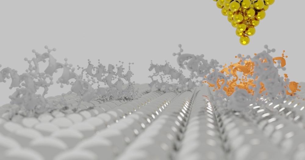
This article discusses the recent advancements in post-scanning image processing of high-speed atomic force microscopy (HS-AFM) for ultrahigh-resolution imaging of complex biomolecules.

Image Credit: sanjaya viraj bandara/Shutterstock.com
The Complexity of Imaging Biomolecules
Analyzing structures and physicochemical properties of biomolecules is a major field of research in biotechnology. The four major types of biological macromolecules namely, carbohydrate, proteins, nucleic acids, and lipids have complex and dynamic conformal states, among which proteins have the most diverse shapes, sizes, and structures.
Traditional electron microscopes (EMs) are suitable for high-resolution imaging of most molecular structures up to a subnanometer scale. However, biomolecules are often susceptible to electron bombardment. Moreover, EMs can only be operated in vacuum conditions, which require reconstructed or isolated macromolecular assemblies, away from their original environmental conditions.
These isolated biomolecules can be in a different conformal state than the original one. Several recent studies have used cryo-EM and cryogenic X-ray crystallography, which are based on the solidification of both biomolecules and a liquid medium. However, they cannot give a real-time image of a biomolecule dynamic conformal state due to changes in temperature, pressure, molecular forces, and buffer exchanges.
Meanwhile, AFM has become a complementary solution to EM in the field of analyzing biomolecules. AFM is a widely used instrument in material science for scanning physical properties and localized forces of 2D surfaces such as topography, magnetic forces, and electrostatic forces through direct physical interaction. Additionally, it can operate under vacuum, air, and a liquid medium with little or no requirement of sample preparation. However, there are still some challenges for HS-AFM in mapping biomolecules. They include the limited sharpness of the AFM probe tip and the sponginess of the molecular chains of the biomolecules in a liquid medium.
The New Localization AFM Technique
A new localization AFM (LAFM) technique has been proposed by Heath et al. to overcome the above-mentioned physical limitations of AFM using a post-probing image processing method. The researchers have explained this method with an analogy, in which a basketball (resembling the AFM probe tip) is dropped on a field containing a large number of ping pong balls (resembling molecular blocks of the biomolecule) randomly hanging in the air (i.e the mean position of molecule blocks). The height of bouncing each ping pong ball will be a measure of their mean position.
The LAFM technique expands the scanned image using the bicubic interpolation method that precisely measures the local maxima (i.e., peak positions) of isolated signals of several closely situated molecule blocks from the images of the sample. Merging of these local maxima in a single frame using a localization algorithm results in a resolution higher than the initial pixel sampling. However, this method is best suited for flat samples without nearby higher features. This provides an equal reference plane to all molecule blocks on a single frame.
Testing the Effectiveness of LAFM
The team first used the LAFM technique for imaging features on the surface of aquaporin-Z (AqpZ) tetrameric channels, a protein that carries water in cell membranes, which look like single protruding loops. They were able to detect hidden structural features with a spacing of 2.6 Å, which was much smaller than the previously recorded value (i.e., 11 Å) using other similar methods including that obtained from the raw data of Nyquist frequency (1/(6.6 Å)). The obtained resolutions of amino acid proteins using LAFM were 4.0 Å, 5.1 Å, and 4.5 Å for AqpZ, A5, and A5 P13W-G14W, respectively.
More from AZoM: Utilization of Multi-Frequency Magnetic Atomic Force Microscopy
Furthermore, to study the effectiveness of LAFM for biomolecules in buffer liquid medium, the researchers used CLC-ec1, a Cl−/H+ antiporter from E. coli. The high-resolution dynamic real-time mapping showed that the dimeric proteins protruding from its membrane exhibited a pH-dependent change of the conformal state.
Conclusions
LAFM is a promising algorithm-based post-probing image processing method to obtain high-resolution AFM mapping that helps overcome the physical limitations of traditional HS-AFM in mapping very small biomolecule chains. LAFM offers a resolution beyond the tip radius limit even for biomolecules in a non-rigid state, unlike cryo-EM.
It is also a suitable alternative to EMs, which cannot map these molecules in their original environmental conditions or a dynamic situation. LAFM mapping can be performed for many molecules in one or several frames or a single molecule over time and could soon become the standard characterization method for complex biomolecules.
Reference and Further Reading
Heath, G., Kots, E., Robertson, J., Lansky, S., Khelashvili, G., Weinstein, H., Scheuring, S., Localization atomic force microscopy. Nature, 594, 385–390 (2021). https://www.nature.com/articles/s41586-021-03551-x
Disclaimer: The views expressed here are those of the author expressed in their private capacity and do not necessarily represent the views of AZoM.com Limited T/A AZoNetwork the owner and operator of this website. This disclaimer forms part of the Terms and conditions of use of this website.