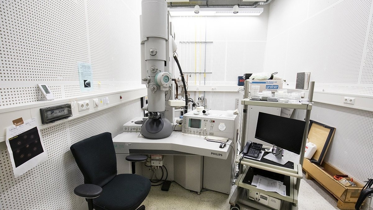
Transmission Electron Microscopy (TEM) enables scientists to examine the structure of materials with far greater resolution than an optical microscope. However, atomic resolution can only be achieved consistently with aberration correction.

Image Credit: Dectris Ltd
This year, the electron microscopy community marks the 25th anniversary of the release of the first aberration-corrected transmission electron microscope. To celebrate the occasion, this article presents an overview of the top five leading publications on the subject.
In the 1930s, shortly after Ernst Ruska invented TEM, researchers were investigating methods for correcting electron lenses from the naturally existing spherical aberration (Cs).
However, Cs-correction for (S)TEM microscopes was only realized in the 1990s, paving the way for sub-ångström resolutions. The credit for this breakthrough goes to Harald Rose, Max Haider, Knut Urban, and Ondrej Krivanek – along with many of their collaborators.
The research on aberration correction has produced many highlights. To commemorate its 25-year history, this article summarizes the top five publications on the subject.
1. Aberration-Corrected Scanning Transmission Electron Microscopy for Atomic-Resolution Studies of Functional Oxides
I. MacLaren, Q. M. Ramasse, International Materials Reviews, 59:3, 115-131
This paper is a concise history of the technology, helping readers refresh their STEM knowledge and look into the applications of aberration-corrected STEM for functional oxides. The authors also discuss possible future directions in AC-STEM.
2. High-Resolution Imaging with an Aberration-Corrected Transmission Electron Microscope
M. Lentzen, B. Jahnen, C. L. Jia, A. Thust, K. Tillmann, K. Urban, Ultramicroscopy Volume 92, Issues 3–4, August 2002, Pages 233-242
The authors studied the ramifications of the variable spherical aberration and proposed novel imaging modes. The research article also examined new applications of the instrument in semiconductor heterostructures and ceramic grain boundaries.
3. Toward Sub-Å Electron Beams
O. L. Krivanek, N. Dellby, A. R. Lupini, Ultramicroscopy Volume 78, Issues 1–4, June 1999, Pages 1-11
In this breakthrough study, the authors described the construction of a CS corrector that offers special attention to the impact of instabilities and shows excellent results.
4. Atomic-Resolution Protein Structure Determination by cryo-EM
K. M. Yip, N. Fischer, E. Paknia, A. Chari, H. Stark, Nature volume 587, pages157–161 (2020)
In this paper, the researchers argue that “the direct visualization of atom positions is crucial for comprehending the workings of protein-catalyzed chemical reactions”. For the first time, this study presented a 1.25 Å-resolution structure of apoferritin derived using cryo-EM with a newly constructed aberration-corrected electron microscope.
5. Real-Space Visualization of Intrinsic Magnetic Fields of an Antiferromagnet
Y. Kohno, T. Seki, S. D. Findlay, Y. Ikuhara, N. Shibata, Nature volume 602, pages234–239 (2022)
This paper examines an enhanced electron optics design for high-resolution EM imaging of magnetic samples. The authors present a real-space visualization of the magnetic field distribution inside antiferromagnetic haematite (α-Fe2O3) using atomic-resolution DPC STEM in a magnetic-field-free environment.

This information has been sourced, reviewed and adapted from materials provided by Dectris Ltd.
For more information on this source, please visit Dectris Ltd.