Electron Energy Loss Spectroscopy (EELS) measures how much energy electrons lose while interacting with a sample. These findings are then utilized to characterize a broad range of materials.
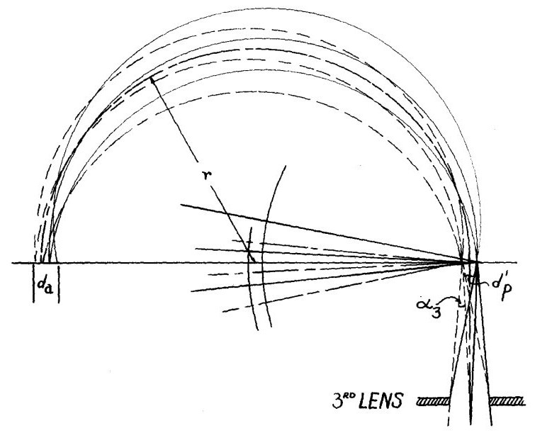
A diagram showing the ray path in the 180° magnetic deflection type of electron velocity analyzer incorporating a focusing lens. Source: Microanalysis by Means of Electrons
J. Hillier and R.F. Baker first developed this technique in the 1940s. However, it took more than 50 years and substantial advancement in microscopy equipment to claim its proper place today.
A great deal has happened in the nearly century-long existence of EELS. This article presents some of the most fascinating publications on the subject selected after a thorough literature review.
The electron energy loss (EEL) spectrum can be considered a material’s fingerprint. It spans an energy range of millielectronvolts to kiloelectronvolts and comprises data regarding the nature of chemical bonds, dielectric properties, band structures, structural configurations, electrical properties, and thermal characteristics of the sample.
Nowadays, the approach has improved to the point where many quasiparticles routinely leave their footprints on the EEL spectra, and much effort is being put into improving the visibility of these features.
Researchers have been refining the technology for nearly a century, exploiting advances in electron optics and detection technologies. For instance, hybrid-pixel direct electron detectors work particularly well when paired with EEL spectrometers, allowing more speed and experimental versatility.
This article reviews the vast body of literature on the subject, picking some of the early essential works on technique development, some contemporary articles that explain cutting-edge instrumentation, and one exotic publication with a "precious" turn-up.
1. Parallel Detection Electron Spectrometer Using Quadrupole Lenses
O.L. Krivanek, C.C. Ahn, R.B. Keeney, 1987
Ondrej Krivanek, a recent Kavli Prize recipient, made substantial contributions to the advancement of electron energy loss spectrometers while working at Gatan, ensuring the decade-long popularity of post-column spectrometers.
This paper discusses how adding three quadrupole lenses to a post-column spectrometer, a linear arrangement of photodiodes with a YAG scintillator, and a computer-controlled data transfer interface significantly improved the instrument’s operational efficiency and flexibility.
When photographic films were still a viable option, this method permitted acquisition times of fractions of a second, which many Ph.D. students greatly welcomed.
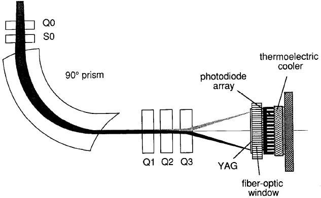
Schematic diagram of the parallel detection system showing the magnetic sector, the pre-sector focussing coils, the post-spectrometer lens assembly, and the photodiode detection system. Image Credit: Dectris Ltd
2. Developments in EELS Instrumentation for Spectroscopy and Imaging
O.L. Krivanek, A.J. Gubbens, N. Dellby, 1991
An EEL spectrum undoubtedly provides a wealth of information, but the goal when using a microscope is to concentrate on images. By 1991, parallel EELS was a well-established technology, thus Gatan focused its R&D efforts on merging spectroscopic and imaging data.
This paper outlines the concepts behind their first energy filter (GIF, in the future). It enabled TEM and STEM modes to pick an energy window and generate images utilizing only those electrons with a defined interaction with the sample. Suddenly, elemental mapping became accessible, and (S)TEM images had to be printed in colors.
The requirement to correct multiple orders of aberrations to generate undistorted and sharp images significantly increased the complexity of the electron-optics design and the algorithms used to optimize the spectrometer alignments.
Fortunately, the user was supported by powerful software that did its best to improve the spectrometer's performance.
3. Test and Characterization of A New Post-Column Imaging Energy Filter
F. Kahl, V. Gerheim, M. Linck, et al., 2019
From an electron optical standpoint, designing an energy filter involves identical analytical and mathematical procedures as designing a multipole aberration corrector.
Given their extensive expertise in the sector and excellent academic background, the researchers at CEOS GmbH set out to build a novel post-column imaging filter to enhance the performance of commercially available products.
This project's important aims were stability, reproducibility, and simplicity of use, as demonstrated in this 2019 article. The article fully characterizes the CEOS Energy-Filtering and Imaging Device (CEFID).
4. Single-Particle cryoEM at Atomic Resolution
T. Nakane, A. Kotecha, A. Sente, et al., 2020
Imaging filters serve Materials Science and Life Sciences, although this approach has only recently overcome the osmotic barrier that divides the two communities.
In thick amorphous substrates, zero-loss energy-loss filtering may reduce image degradation caused by inelastic scattering. This, combined with other enhancements in their setup, enabled the group from the MRC Laboratory of Molecular Biology in Cambridge and partners to push the limits for single-particle cryoEM.
Employing a cold field emission gun, a next-generation direct electron camera, and a Selectris energy filter created by Thermo Fisher Scientific, the team successfully reconstructed Apoferritin with an impressive resolution of 1.22 Å, building on the concepts described in the previous article.
5. EELS Femtosecond Resolved in 4D Ultrafast Electron Microscopy
F. Carbone, B. Barwick, O. Kwon, et al., 2009
Nature is unstable: materials undergo phase changes, chemical processes are not static, and heat and sound disperse. To truly examine physical systems in their natural condition, time-resolved approaches are required, in which samples are forced into excited states by an impulsive stimulus and then studied while relaxing.
In the research facilities of Nobel Prize winner Ahmed Zewail, known as "Femtoland," a couple of microscopes were modified to incorporate ultrafast lasers to investigate reversible physical processes stroboscopically, and multiple standard TEM methods were extended to the fourth dimension.
Carbone et al. employed ultrafast EELS to investigate the behavior of graphite samples after a brief laser pulse.
Measuring the intensity of the plasmon peak corresponding to the interaction across layers reveals the signature of an ultrafast phase transition from graphite to diamond, the most well-known carbon allotropes.

This information has been sourced, reviewed and adapted from materials provided by Dectris Ltd.
For more information on this source, please visit Dectris Ltd.