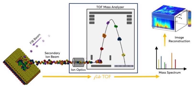
By adding focused ion beam secondary ion mass spectrometry (FIB-SIMS) abilities to FIB-SEM microscopes, the fibTOF enables 3D sensitive imaging of chemicals with nanometer resolution.
- Allows sensitive detection of light elements, like hydrogen, fluorine, lithium and boron
- Enables 3D chemical imaging of all elements with depth profiling resolution of less than 10 nm and lateral resolution of less than 50 nm
- Can be used with leading commercial FIB-SEM microscopes without affecting the quality of images
- Allows unequivocal elemental identification with an increased mass resolving power
- Enables isotopic imaging for experiments to analyze diffusion, transport, or reaction mechanisms
Advancing FIB-SIMS Without Compromise
The fibTOF Adds 3D Chemical Imaging to FIB-SEM Microscopes
Secondary ion mass spectrometry, or SIMS, is a proven method where an energetic ion beam is used for sample sputtering, causing the ejection of both charged particles (secondary ions) and neutral particles.
A mass analyzer is used to measure the sputtered ions, offering chemical data about the specimen. When an appropriately fine primary ion beam is scanned over the specimen, the SIMS technique offers a superior lateral resolution and a high depth resolution (the secondary ions come only from the sample surface).

Image Credit: TOFWERK
By adding the fibTOF capabilities to a FIB-SEM microscope, SIMS measurements can be made with exceptional imaging performance without affecting EDX/SEM measurements. The time-of-flight mass analyzer of the fibTOF invariably achieves a complete mass spectrum, making it possible to visualize any target ions during the post-processing of data.
Specifications of fibTOF
The fibTOF can be used with leading commercially available FIB-SEM microscopes without affecting the image quality.
Source: TOFWERK
Mass Resolving Power
M/ΔM FWHM |
Mass Range (Th) |
Limit of Detection |
Lateral Spatial Resolution* |
Depth Resolution* |
| >700 |
1 – 500 |
ppm |
50 nm |
10 nm |
*Based on the performance of the focused ion beam.