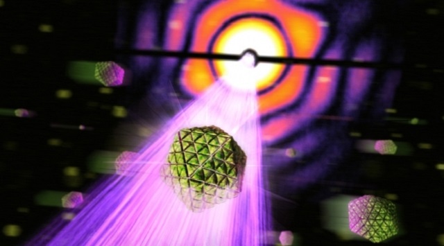
 Carboxysomes are sprayed across the very intense beam of an X-ray laser. Whenever a particle is hit a diffraction pattern is produced from which the structure of the particle can be calculated. Photograph: SLAC National Accelerator Laboratory
A research team from Uppsala University has developed an imaging method to study biological particle structures using X-ray lasers. They used carboxysome – a tiny cellular organelle in photosynthetic bacteria to test the method. The results showed that the technique provides an in-depth knowledge in understanding the structure of smaller organisms through 3D imaging.
Carboxysomes are tiny structures carrying protein molecules for incorporating carbon into biomolecules. They play a major role in global carbon fixation. The researchers, at the SLAC National Accelerator Laboratory, sprayed carboxysomes across the LCLS X-ray laser beam with the help of a special injector producing a extremely thin particle stream. The structure of carboxysomes scattering the short and ultra-bright x-rays were analyzed to determine the structure of the organelles. The extreme brightness of the x-rays enabled the researchers to reconstruct samples without crystallization.
Around 70,000 scattering patterns of individual particles in the sample were collected in 12 minutes. The results showed significant variation in the size of the sample in addition to returning an icosahedral shape of the structure. Using X-ray lasers, it is possible to obtain the image of whole living cells at an unmatched resolution.
The researchers successfully reconstructed samples such that the results show details as small as 18nm. This paves the way for imaging large pathogenic viruses such as herpes, influenza and HIV that are in similar size of carboxysome. In addition, the size distribution of the carboxysomes before and after the experiment remained the same, which denoted that the imaged organelles remain intact.
As the strong X-ray pulses tend to destroy the sample, the diffraction pattern can be precisely obtained prior to disintegration, using “diffraction-before-destruction” method. This single-particle imaging technique will come in existence by 2017 after opening a new facility at the European XFEL.
These advances lay the foundations for accurate, high-throughput structure determination by flash-diffractive imaging and offer a means to study structure and structural heterogeneity in biology and elsewhere.
Professor Janos Hajdu, Lead Author of the paper
References