Oct 27 2017
The first ever atomic-scale images of dendrites (i.e. digitated growths), which have the ability to penetrate the barrier in between battery compartments and to trigger fires or short circuits, have been captured by researchers from Stanford University and the Department of Energy’s SLAC National Accelerator Laboratory.
Dendrites and the challenges they pose are a major obstacle in designing innovative kind of batteries with the potential to store greater energy such that cell phones, electric cars, laptops, and other devices can be operated for a longer period of time before being recharged.
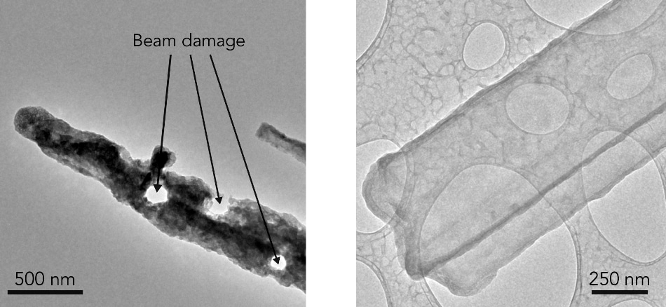
 Left: In this room-temperature TEM image, exposure to air has corroded a lithium metal dendrite and the electron beam has melted holes in it. Right: In contrast, a cryo-EM image of a dendrite shows that freezing has preserved its original state, revealing that it’s a crystalline nanowire with well-defined facets. CREDIT: Y. Li et al., Science.
This is the first research to investigate the reactions inside batteries by adopting cryo-electron microscopy (i.e. cryo-EM), a method that was awarded with the 2017 Nobel Prize in chemistry for its potential to image flash-frozen, delicate proteins and other “biological machines” at the atomic level.
The new images demonstrate that each lithium metal dendrite is of the form of a long, aesthetically formed six-sided crystal, in contrast to the pitted, irregular shape illustrated in earlier electron microscope images. According to the researchers, the potential to observe details at this level for the first time by adopting cryo-EM will offer researchers with a robust tool for acquiring an in-depth knowledge on the working of batteries and their components at the most basic level and for analyzing the reason behind the occasional failure of high-energy batteries used in cell phones, laptops, electric cars, and airplanes. The results of the study have been published in the journal Science on October 26, 2017.
This is super exciting and opens up amazing opportunities, with cryo-EM, you can look at a material that’s fragile and chemically unstable and you can preserve its pristine state—what it looks like in a real battery—and look at it under high resolution, this includes all kinds of battery materials. The lithium metal we studied here is just one example, but it’s an exciting and very challenging one.
Yi Cui, professor at SLAC and Stanford and investigator with SIMES
Fickle Fingers of Failure
Cui’s laboratory is one of various developing approaches—such as creating a “smart” battery with the ability to automatically turn off upon sensing the invasion of dendrites into the barrier between the chambers of the battery, or adding chemicals to the electrolyte to prevent their growth—to avoid damages caused by dendrites.
However, to date, researchers have been largely unsuccessful in acquiring atomic-level images of dendrites or any other sensitive battery parts. The desired method (i.e. TEM or transmission electron microscopy) was found to be very abrasive for several materials, such as lithium metal.
“TEM sample preparation is carried out in air, but lithium metal corrodes very quickly in air,” stated Yuzhang Li, a Stanford graduate student who headed the study along with Yanbin Li, a fellow graduate student. “Every time we tried to view lithium metal at high magnification with an electron microscope the electrons would drill holes in the dendrite or even melt it altogether.”
“It’s like focusing sunlight onto a leaf with a magnifying glass. But if you cool the leaf at the same time you focus the light on it, the heat will be dissipated and the leaf will be unharmed. That’s what we do with cryo-EM. When it comes to imaging these battery materials, the difference is very stark.”
Batteries Take a Freezing Dip
The cryo-EM technique involves flash-freezing the samples by immersing them into liquid nitrogen, and subsequently slicing them for investigation under the microscope. It is possible to freeze an entire coin-cell battery at a specific point in time during its charge-discharge cycle, isolate the component of interest, and observe the reactions inside that component at the atomic scale. It is also feasible to produce a stop-action video of battery activity by linking images captured at different time points in the cycle.
To perform the research, the scientists used a cryo-EM instrument from Stanford School of Medicine to investigate thousands of dendrites of lithium metal that were exposed to different electrolytes. Apart from observing the metallic part of the dendrite, they also observed a coating known as SEI (i.e. solid electrolyte interphase) that is developed when the dendrite interacts with the surrounding electrolyte. The same coating is also formed on metal electrodes during the charging and discharging cycle of the battery. Arresting the formation of this coating and regulating its stability are inevitable for effective battery functioning.
Astonishingly, they found that the dendrites are faceted, crystalline nanowires that opt to grow in specific directions. Although certain dendrites developed kinks when they grew, their crystal structure fascinatingly stayed unaltered despite these kinks.
A Zoom Lens for Atoms
The team zoomed in and tried an alternative method for observing the manner in which electrons escaped from the atoms in the dendrite, thereby disclosing the locations of single atoms in the crystal as well as its SEI coating. Upon adding a chemical, usually used to enhance battery performance, the SEI coating’s atomic structure was shown to be well-ordered, and the researchers believe that this might help explain the reason for the working of the additive.
We were really excited. This was the first time we were able to get such detailed images of a dendrite, and we also saw the nanostructure of the SEI layer for the first time, this tool can help us understand what different electrolytes do and why certain ones work better than others.
Yanbin Li, a graduate student who headed the study
According to the team, in the future, they hope to concentrate on acquiring in-depth knowledge of the structure and chemistry of the SEI coating.
Researchers from the Stanford School of Medicine, ShanghaiTech University and University of Siegen also contributed to this study. The DOE Office of Vehicle Technologies supported the study under the Battery Materials Research Program and Battery 500 Consortium.
Cryo-EM | Close-up image of a lithium metal dendrite
This video demonstrates how cryo-EM is able to get a close-up image of a lithium metal dendrite when other methods fail. In the first half, a dendrite curls up and melts when hit with an electron beam during transmission electron microscopy, or TEM, which takes place at room temperature. The second half zooms in on a flash-frozen dendrite – magnified about 400,000 times, so it nearly fills the frame – during cryo-EM imaging. Freezing protects it from the electron beam so scientists can see its intact structure for the first time, down to an atomic level. The solid electrolyte interphase (SEI) layer that coats the dendrite and its crystalline components are also clearly visible along its right edge. (Yuzhang Li/Stanford University)