Feb 10 2020
During the production of cosmetics and detergents, it is important to control the structure of proteins. But so far, there has been no clear understanding of how both proteins and soap molecules work together to alter the structure of proteins.
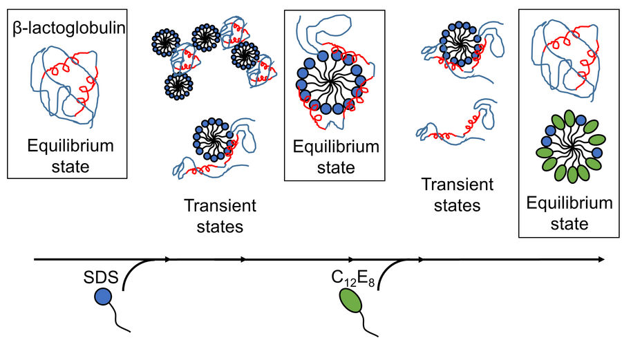
 Results published by AU researchers reveal that surfactant-mediated unfolding and refolding of proteins are complex processes with several structures present, and rearrangements occur on time scales from sub-milliseconds to minutes. Image Credit: Reproduced with permission from the Royal Society of Chemistry.
Scientists at Aarhus University have successfully produced a comprehensive picture of how soap molecules are able to refold and unfold the proteins on the millisecond timescale.
Figuring out the interactions between soap molecules (surfactants) and proteins has traditionally been significant for the industry, specifically within cosmetics and detergents.
It is known that sodium dodecyl sulfate (SDS)—an anionic surfactant—unfolds globular proteins, whereas octaethylene glycol monododecyl ether (C12E8)—a non-ionic surfactant—does the reverse, that is, it helps proteins to refold into a shape.
If washing powders had to work efficiently, then it should be ensured that the surfactants do not alter the structure of proteins or enzymes. This is because any changes in the structure of the enzymes destroy their potential to remove dirt or break down stains.
A majority of the washing powders contain a combination of surfactants that enable the enzymes to stay active. Moreover, certain biotechnologies tend to manipulate the surfactants along with proteins.
Generally, membrane proteins are present in the cell membrane. To extract these membrane proteins from this setting for various analyses, they need to be solubilized by the surfactant. This surfactant should be sufficiently “gentle” and only enclose the membrane-inserted portion of the proteins, so that their structure is not disturbed.
On the other hand, when the molecular weight of proteins is being characterized in the laboratory, one typical method is to unfold these proteins by SDS, which happens to be the aggressive negatively charged surfactant, and track how these proteins move in a polymer gel within an electric field. But this method works only when the surfactant fully unfolds the proteins and damages their structure.
A debate is still going on about which kind of interactions between the surfactant and the protein is most significant. Is it the electrostatic interactions that occur between the protein and the surfactant charges, or is it only the characteristics of the interface of the aggregates (micelles) that the surfactants form in water, which account for protein unfolding?
Despite a thorough analysis of the unfolding processes at the protein level, a clear picture of the communication between surfactant and protein is not available in these processes. This lack of understanding has been addressed in the present work by utilizing the globular protein β-lactoglobulin, or bLG, as a model protein.
The Right Combination of Experimental Techniques
A better understanding of the refolding and unfolding of proteins was achieved by plotting the numerous steps of protein-surfactant interactions as a function of time.
At first, bLG—the model protein—was combined with the anionic surfactant SDS and, at the same time, the time evolution of the development of complexes between surfactant and protein molecules was tracked on the time scale ranging from milliseconds to minutes.
This method allowed the scientists to establish the structure of the emerging complexes. The team then plotted the time course of the refolding process when C12E8, a non-charged surfactant, was introduced to a sample comprising complexes of protein and SDS.
To visualize the way the protein reassembles during the course of the refolding and unfolding process caused by surfactants, complementary spectroscopic methods like tryptophan fluorescence and Circular Dichroism were employed together with time-resolved Small-angle X-ray scattering, or SAXS.
While variations in the bLG structure were tracked by both tryptophan fluorescence and Circular Dichroism, variations in the total shape of the complexes of protein and surfactant were monitored by synchrotron SAXS. Earlier, such kinds of combined techniques have never been used to analyze these processes.
Complex Processes Lasting Milliseconds to Minutes
Protein unfolding by SDS was a uniform process, in which all the molecules of proteins follow the same path of unfolding. The SDS complexes, or micelles, directly attack the protein molecules and then slowly unfold the protein so that it creates a shell over the SDS micelle. The refolding process begins when C12E8 micelles form mixed SDS-C12E8 micelles by “sucking out” the SDS from the protein-SDS complex.
But the actual refolding process appears to follow a number of paths, as numerous structures were observed to form simultaneously, such as mixed micelles of C12E8 and SDS, protein-surfactant complexes (perhaps comprising both C12E8 and SDS), properly folded proteins, and “naked” proteins that unfolded just like long polymeric chains.
The experiment made it possible to track the inter-conversion between these species, so that the type of processes that are fast and the ones that are slow can be determined.
The folded protein is likely to form from the naked unfolded proteins (quickly) and also from the complexes of protein and surfactants (more gradually). Hence, the most optimal way where surfactants can assist in the protein folding process is to essentially get out of the way and allow the protein to trace its own way back to the folded state.
The outcomes have given a better understanding of the structural variations that take place at the protein–surfactant level. The results also demonstrated that refolding and unfolding of proteins mediated by surfactants are intricate processes of rearrangements that take place on time scales from less than milliseconds to minutes and also involve a close association between proteins and surfactant complexes.
The Independent Research Fund Denmark funded the study. The study was performed by scientists from Interdisciplinary Nanoscience Center (iNANO) and Department of Chemistry, and Department of Molecular Biology and Genetics at Aarhus University, in association with scientists from ESRF—The European Synchrotron in Grenoble (France).
Professor Jan Skov Pedersen (iNANO and Department of Chemistry, Aarhus University) and Professor Daniel E. Otzen (iNANO and Department of Molecular Biology and Genetics, Aarhus University) were in charge of the research group.