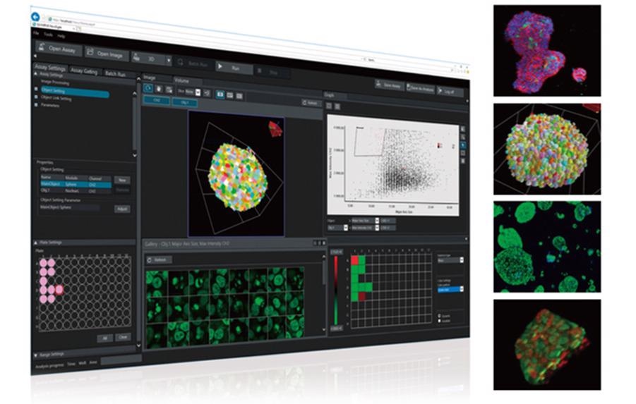
Previously only available in the U.S., Olympus is launching NoviSight 3D analysis software globally. For life science research that relies on cell analysis, the information provided by NoviSight software can improve the entire experiment process.
 Olympus NoviSight Software. Image Credit: Olympus Scientific Solutions Americas NDT
The need for 3D cell analysis
As opposed to conventional 2D cell analysis, the use of 3D cell models that more accurately simulate a living body can speed up experiments. For example, using 3D cultured cell models, such as spheroids and organoids, researchers can analyze the effects and toxicity of new drugs in a bioenvironment similar to a living human body. This approach improves the accuracy of results at the preliminary stage of drug discovery.
Insightful data, intelligent analysis
Olympus has combined 3D imaging technology with powerful algorithms to create software capable of analyzing a whole cell model in three dimensions. Used with Olympus confocal laser scanning microscopes, such as the FLUOVIEW FV3000 system, NoviSight 3D cell analysis software provides images of cell clusters down to the nuclei. The software’s True 3D technology uses multiple microplate images to provide accurate morphology data and the ability to quantitatively analyze the effect of medications, including growth suppression and cell survival rates. A range of parameters can be easily and precisely measured, enabling researchers to count the number of cells that have suppressed growth, proliferated or have died. NoviSight 3D cell analysis software also makes it easy to compare the effects of different medications at various concentrations.
NoviSight software can facilitate and accelerate the interpretation and validation process. Results, including recognition, analysis and statistics, are displayed on a single screen. Users can view the quantitative data as a scatterplot, heat map or graph, and clicking on a point in the graphical display automatically opens the corresponding image. It is easy to switch between 2D and 3D views of the sample, and data are easily exported as a CSV or FCS file for further analysis.
With the advancement of cell culture technology, working with 3D cell culture samples is increasingly common. NoviSight software’s powerful capabilities provide the critical tools needed to analyze 3D cell cultures to potentially save researchers time and reduce costs.
To learn more about NoviSight software, visit: Olympus-LifeScience.com.