 By Surbhi JainReviewed by Susha Cheriyedath, M.Sc.May 5 2022
In an article recently published in the open-access journal Polymers, researchers discussed the development of interfaces based on laser-structured arrays of carbon nanotubes with albumin for electrical stimulation of heart cell growth.

Study: Interfaces Based on Laser-Structured Arrays of Carbon Nanotubes with Albumin for Electrical Stimulation of Heart Cell Growth. Image Credit: picmedical/Shutterstock.com
Background
Biomedical research is grappling with the production of biocompatible materials with certain qualities. Carbon nanotubes (CNT) are a one-of-a-kind material that allow for large increases in conductivity while maintaining biocompatibility. The treatment of cardiovascular diseases (CVD), which are currently the leading cause of death in the world, is one of the key uses of electrically conductive nanointerfaces in biomedicine.
At the same time, coating multi-walled carbon nanotubes (MWCNT) arrays with suitable biocompatible polymers can promote cell survival and facilitate a more physiological transition to the soft surface of the human tissues from the hard surface of the MWCNTs. Albumin, a key transport protein present in the blood that supplies numerous nutrients to cells, is one ideal polymer for this purpose.
Nanotubes create contact with each other under the influence of laser light, forming an electrically conductive network. It is, however, incredibly difficult to create a homogeneous liquid dispersion containing uniformly distributed carbon nanotubes. In this context, approaches including the usage of initial nanotubes will be appealing for generating planar nanocomposites with an evenly distributed system of nanotubes.

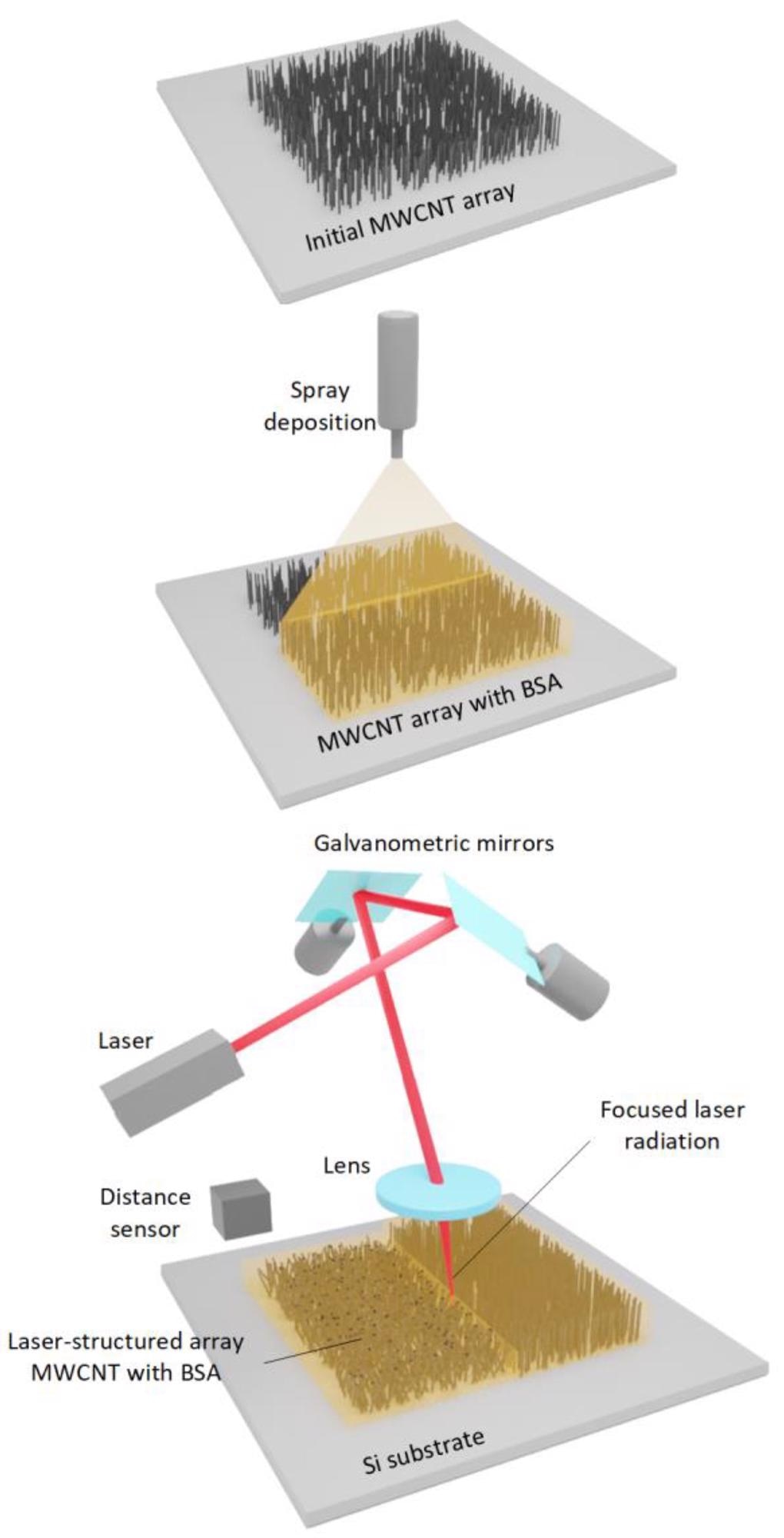
Manufacturing process of electrically conductive interfaces. Image Credit: Fabri, G et al., Polymers
About the Study
In this study, the authors discussed the development of nanostructured arrays of MWCNT in a biopolymer (albumin) layer for enhanced biocompatibility. By using a deposition method, a layer of liquid albumin dispersion was sprayed on manufactured MWCNT arrays. Nanosecond pulsed laser radiation mapping in the near-IR spectral range with λ= 1064 nm was used to create these nanostructures.
The team suggested a method for the formation of electrically conductive materials derived from vertical MWCNT arrays densely distributed all over the silicon substrate area. Vertical arrays were coated with albumin and exposed to laser light to generate biopolymer materials having connections between nanotubes.
The researchers used energy-dispersive spectroscopy, electron microscopy, and Raman spectroscopy to examine the morphology of the prepared materials. The influence of the energy density of laser radiation on nanomaterial shape and electrical conductivity was also discussed. The suitability of the prepared nanoparticles as interfaces in the electrical stimulation for the growth of cardiac and connective tissue cells was demonstrated using the established installation.

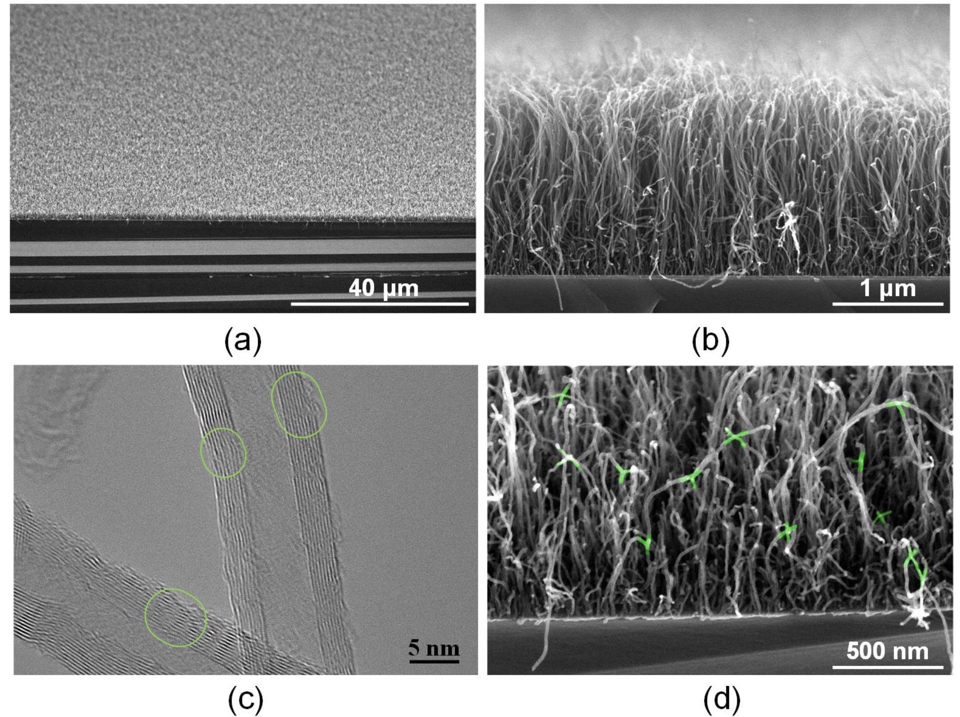
SEM (a,b) and TEM (c) images of MWCNT arrays on Si substrate before and after laser exposure with an energy density of 0.013 J/cm2 (d). Green ovals show transition regions in which there is a higher number of defects in the area compared to that in the defect-free part of the nanotube framework. Nanotube binding regions are highlighted in green. Image Credit: Fabri, G et al., Polymers
Observations
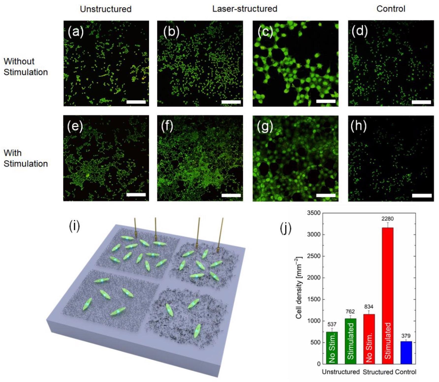
Cell densities of 500 mm-2 were obtained in the control samples, which were equivalent to typical values for fibroblast-coated substrates. In comparison to the control, the density of cells increased for both non-stimulated and stimulated unstructured arrays and reached a value of 740 mm-2 and 2050 mm-2, respectively. Electrical conductivity incremented by more than two times when subjected to a laser with an energy density of 0.015 J/cm2 and reached a value of 215.8 ± 10 S/m. The electrical conductivity was reduced after the radiation energy density was increased to 0.017 J/cm2.
The energy density of 0.015 J/cm2 was determined to be sufficient for the MWCNT structure. The structuring effect happened during the production of C–C bonds in the albumin matrix, which occurred simultaneously with the formation of a cellular structure of nanotubes. It resulted in a decrease in nanotube defectiveness, as measured by Raman spectroscopy. Furthermore, laser structuring increased the electrical conductivity of MWCNT arrays to 215.8 ± 10 S/m containing albumin by more than threefold. Cells on the interfaces with the system based on a culture plate were electrically stimulated, resulting in increased cell proliferation.
The process of plasma-enhanced chemical vapor deposition on a silicon substrate from the gas phase was used to transform biocompatible nanostructures from the arrays of MWCNTs. Using nanosecond pulsed laser energy in the near-infrared region of the spectrum, nanostructures were generated by modifying the shape of the MWCNT array present in the albumin matrix at the same time as the nanotubes were bonded to each other. It was feasible to reduce the defectiveness of the tubes by using laser therapy, as evidenced by Raman spectroscopy data.
The optimal ratio of nanotubes and albumin in the samples, along with the formation of junctions between nanotubes and a cellular structure, was identified using a scanning electron microscope (SEM) at a radiation energy density value of 0.015 J/cm2. When compared to the initial MWCNT arrays having a conductivity of 100.7 ± 2 S/m, the proposed structure had a more than threefold increase in electrical conductivity and obtained a value of 215.8 ± 10 S/m.

Examples of fluorescence microscope images of fibroblast cell cultures on Si substrates 24 h in vitro with unstructured (a,e) and laser-structured (b,c,f,g) MWCNT arrays with BSA immobilized with (e–g) and without (a–c) electrical stimulation and control samples (d,h). The scale bar for (a,b,d–f,h) is 200 µm, for (c,g) it is 50 µm. Schematic of fibroblasts grown on Si substrates with various conditions used in the experiment (i). The values on the bars in (j) represent the total number of live cells averaged over several areas of size 850 µm × 850 µm (0.7225 mm2). The results from three images on three different samples for each type of the condition were averaged for the statistics; n = 9. Image Credit: Fabri, G et al., Polymers
Conclusions
In conclusion, this study discussed the development of a system for the electrical stimulation of cells. The proposed setup was used to stimulate and investigate the effect of electrical stimulation on differentially customized surfaces for fibroblasts and cardiomyocytes. For both types of cell cultures, electrical stimulation resulted in a considerable increase in cell proliferation rate on structured arrays.
The authors suggested that laser-structured MWCNT arrays with biopolymer albumin coatings could be a useful tool for bioelectronics devices, improving cell experiments, and potential biomedical applications such as heart implants and biosensors. They also mentioned that MWCNT laser-structured arrays with biopolymers appear to be a promising material for a wide range of biological applications.
Disclaimer: The views expressed here are those of the author expressed in their private capacity and do not necessarily represent the views of AZoM.com Limited T/A AZoNetwork the owner and operator of this website. This disclaimer forms part of the Terms and conditions of use of this website.
Source:
Fabri, G., Ometto, A., Villani, M., et al. Interfaces Based on Laser-Structured Arrays of Carbon Nanotubes with Albumin for Electrical Stimulation of Heart Cell Growth. Polymers 14(9) 1866 (2022). https://www.mdpi.com/2073-4360/14/9/1866