 By Surbhi JainReviewed by Susha Cheriyedath, M.Sc.Oct 10 2022
In an article recently published in the open-access journal Scientific Reports, researchers discussed the resistance to fracture of variously designed bonded ceramic overlay restorations.

Study: Fracture resistance of bonded ceramic overlay restorations prepared in various designs. Image Credit: 58Ararae/Shutterstock.com
Background
The two main inorganic components of a tooth are dentin and enamel, and each has special qualities that benefit the other. The enamel's firmness safeguards the dentin and pulp tissue, the tooth's underlying important components, from damage while the dentin's flexibility gives the tooth its overall flexibility and helps prevent the enamel from fracturing. There are currently no substitutes for this ideal blend of nature in restoration materials. Therefore, it is always best to keep as much of the original tooth structure as possible.
Dental bonded restorations have been created to conserve as much of the intact tooth structure. The keys to successfully adhering to a ceramic or metal restoration to the native tooth are a good dental adhesive system and resin cement. The intact tooth structure not needed for the retention of the restoration is removed during tooth preparation for a full coverage crown.
For this type of restoration, bondable restorative materials, typically dental ceramics and resin-base materials, as well as a dental adhesive system and resin cement, are advised. The goal is to preserve as much of the natural tooth structure as possible, which must be balanced with the need for the restoration to have a high fracture resistance to chewing pressures. However, there are currently few unambiguous recommendations for conservative tooth preparation before a ceramic overlay.

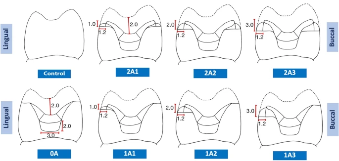
Illustrations of the various preparation designs in this study. Image Credit: Channarong, W et al., Scientific Reports
About the Study
In this study, the authors discussed the fracture resistance of adhesive ceramic overlays with varied designs. Eight groups of 48 upper premolar teeth were created. The variations included contra bevel margins on the buccal and lingual surfaces, shoulder margins on the buccal and lingual surfaces with axial wall heights of 1, 2, or 3 mm, one shoulder margin with an axial wall height of 1, 2, or 3 mm on the lingual surface and one contra bevel margin on the buccal surface, and control of sound teeth. Lithium disilicate ceramic with zirconia reinforcement was used to design and create the overlays, which were then cemented with resin cement.
The team conducted thermocycling and dynamic fatigue tests on the samples to simulate six months of use. Compressive loading was used up till fracture, and the mode of fracture was studied. The majority of fracture patterns included pulp tissue and occurred below the CEJ, and the results demonstrated no statistically significant difference in fracture resistance between designs. The restored teeth's resistance to fracture was statistically coherent with the control's.
The researchers demonstrated that all control fractures occurred above the CEJ and within the dentin. As a result, axial wall heights and margin types had no bearing on this outcome, and overlay restorations were successful in strengthening damaged teeth and granting fracture resistance comparable to sound teeth. This study aimed to evaluate the margin preparation designs and fracture resistance of various axial wall heights for adhesive ceramic overlays. The null hypothesis stated that the fracture resistance of bondable glass ceramic restorations was unaffected by various overlay preparation designs.

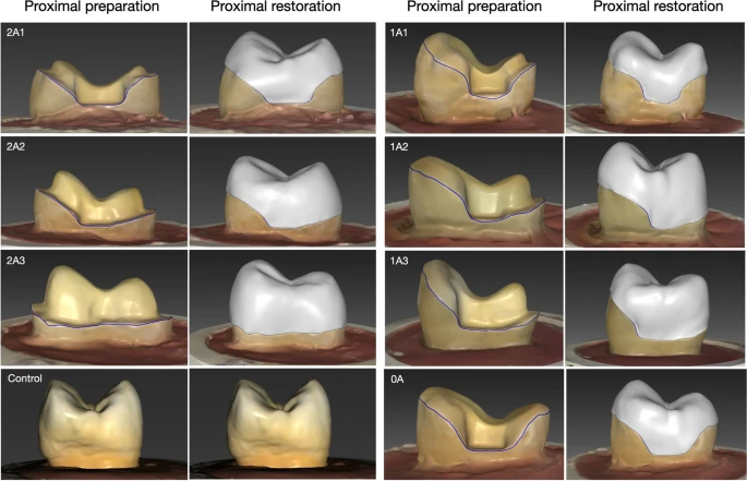
The tooth preparation and the overlay restoration design for each experiment group. Image Credit: Channarong, W et al., Scientific Reports
Observations
There was no statistically significant difference in the fracture resistance of the various ceramic overlay restoration designs examined or between those restorations' fracture resistance and that of the healthy teeth used as the control. As a result, the kind of margin and the length of the axial wall had no effect on the ultimate fracture resistance, and the overlay restorations utilizing the adhesive method were able to reinforce the damaged teeth to the level of fracture resistance equivalent to the sound teeth. Restorations could be bonded to the natural tooth structure without the use of mechanical retention thanks to developments in dental adhesive systems and restorative materials.
When adhesive ceramics were placed over human molar teeth with shoulder margins, there was no statistically significant difference in the fracture resistance of walls with 2-, 3-, and 4-mm heights. In the case of conventional procedures, the adhesive technology could make up for the decreased axial wall height. The A0 group had the lowest fracture resistance, while the 2A3 group had the highest fracture resistance.
This was true even though there was no difference in fracture resistance between the various designs. When the remaining tooth structure needed to be strengthened, as in cases of broken teeth or teeth that had root canal treatments, an axial wall height of at least 2 mm was advised.
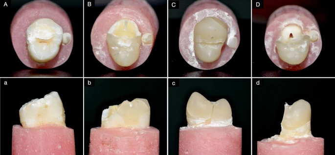
According to the observed fracture patterns, regardless of the design employed, the dominant pattern corresponding to the restoration samples was found between the lingual and buccal cusps, implicated the pulpal tissue, and was located below the CEJ. Although there was no statistically significant difference in the fracture resistance between the sound teeth and the restored teeth, there was a notable distinction in the fracture patterns.
Flexural strength in the range from 130 to 360 ± 60 MPa and fracture toughness in the range from 0.8-1.2 to 2-2.5 MPa m1/2 of the material were both increased by the crystallization and nucleation of the lithium disilicate crystals. The fine grain structure of Li2O-ZrO2-SiO2 crystals was 4–8 times smaller than that of lithium disilicate crystals.

The characteristics of the observed fractures are shown in occlusal view (uppercase letters) and proximal view (lowercase letters). In the control group, the fracture was always within the dentin (A) and it was always above the CEJ (a). In the overlay-restored groups, four of the forty-two specimens involved only the restoration and dentin (B) and three of these unusual specimens were above the CEJ (b). All of the other overlay-restored specimens fractures involved pulp tissue (C and D) below the CEJ (c and d). Roughly half of those specimens fractured across the middle of the tooth (C and c), and in the others a large portion of the tooth fell off (D and d). Image Credit: Channarong, W et al., Scientific Reports
Conclusions
In conclusion, this study discovered that the bondable ceramic overlays' kind of margin and axial wall height had no discernible effect on the restorations' ability to resist fracture. The authors mentioned that depending on the materials being used, different amounts of tooth structure might need to be removed. The elasticity modulus of dental enamel with lithium disilicate ceramic was about 80 GPa.
The team stated that the fracture resistances were identical whether or not the lingual cusps were wrapped around the shoulder borders.
References
Channarong, W., Lohawiboonkij, N., Jaleyasuthumkul, P., et al. Fracture resistance of bonded ceramic overlay restorations prepared in various designs. Scientific Reports, 12, 16599 (2022). https://www.nature.com/articles/s41598-022-21167-7
Disclaimer: The views expressed here are those of the author expressed in their private capacity and do not necessarily represent the views of AZoM.com Limited T/A AZoNetwork the owner and operator of this website. This disclaimer forms part of the Terms and conditions of use of this website.