After only six months from the inauguration of the Joint Electron Microscopy Center at ALBA (JEMCA), this marvel of technology is already feeding published research. In fact, the first paper including data and images obtained with the EM02-METCAM microscope has just been published in the Journal of the American Chemical Society (JACS). And this is set to be the first of many. Recently, the other microscope of the JEMCA center (EM01-Cryo-TEM) also published its first results, proving the outstanding performance of both instruments and its great value for the scientific community.
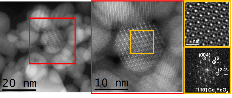
 Atomic resolution aberration corrected HAADF STEM images and corresponding indexed FFT showing that the selected nanoparticle crystallizes along the [110] axis of the Co2FeO4 cubic spinel structure. Image Credit: Jordi Arbiol
The EM02-METCAM (a monochromated and double aberration corrected scanning transmission electron microscope, for the technology-savvies) allows atomic-resolution inspection of the crystalline structure and electronic configuration of materials, something that goes far beyond the mere analysis of their composition. The possibility to study material structures at very small scale is greatly sought after by researchers, not only with the aim of achieving a deeper understanding of chemical and physical phenomena, but also that of engineering novel materials with “custom” (so to speak) properties.
The EM02-METCAM, whose Platform is coordinated by the Catalan Institute of Nanoscience and Nanotechnology (ICN2), is the first microscope of its kind in Spain. With great expectations from the scientists involved in the JEMCA, this powerful tool was immediately put to the test and has gone through a period of commissioning that it has successfully completed.
One of the first studies on which the remarkable properties of the METCAM were tested is the investigation of a family of catalysts –i.e., structures that promote and accelerate certain chemical reactions— that are relevant for application in new types of batteries. Nanoparticles of these catalysts, named “spinel” catalysts for their specific crystal structure, were synthesized by the Functional Nanomaterials group, headed by ICREA Prof. Andreu Cabot, at the Catalonia Institute for Energy Research (IREC) and analysed at the METCAM by researchers from the ICN2 Advanced Electron Nanoscopy Group led by ICREA Prof. Jordi Arbiol. The goal was to shed light on how the geometric configuration, composition, and electronic structure of the nanoparticles under study influence the mechanisms of catalysis.
The new kid on the block proved to be up to the task. The METCAM microscope could easily achieve spatial resolutions below 50 pm –where 1 picometers (pm) is one trillionth of a meter—when operated at an energy of 300 keV and spatial resolutions below 100 pm when operated at 60 keV. The energy resolution achieved at 60 keV when using the monochromator was 13.6 eV.
The atomic-resolution data provided by the METCAM enabled the authors to better understand how these spinel catalysts facilitate reactions and, above all else, to underpin the role of each of their component in the conversion process of polysulfides taking place during charge and discharge of lithium-sulfur batteries. The images obtained with the new equipment were key to resolving the atomic structure of the spinel catalysts. Furthermore, electron energy loss spectroscopy (EELS) information (also provided by the METCAM) was used to study the electronic structure of the surface of the nanoparticles.
Thanks to all these extremely valuable data –combined with the results of other experimental analysis techniques, electrochemical and battery tests, as well as theoretical calculations— the researchers could formulate a comprehensive description of the dependence of spinel catalysts on geometric configuration in the sulfur reduction reaction process. A profound knowledge of the material structure and of the involved reactions represents an essential basis for the rational development of improved catalysts and, ultimately, better-performing batteries.
“The METCAM microscope has definitely exceeded our expectations, consistently achieving resolutions superior to those stated in its technical specifications,” comments Prof. Jordi Arbiol, who is the Scientific Coordinator of the METCAM Platform and co-author of the publication. “The versatility of the multiple detector options, which can be seamlessly combined for both imaging and spectroscopy, empowers us to gain profound insights into materials.” A wealth of achievements and great results are confidently expected for this technological gem, as well as the other cutting-edge microscopes and equipment that constitutes the JEMCA.
The acquisition of the EM02-METCAM was made possible thanks to a collaboration of different research facilities: the Catalan Institute of Nanoscience and Nanotechnology (ICN2) acting as owner of the equipment, the ALBA Synchrotron where the microscope has been installed and is operated complementing the analytical capabilities of the synchrotron beamlines, the Spanish National Research Council (CSIC), the Institute of Materials Science of Barcelona (ICMAB-CSIC), and the Universitat Autònoma de Barcelona (UAB). The project definition phase also included the fundamental support of the Barcelona Institute of Science and Technology (BIST). It was co-financed by the European Regional Development Fund (ERDF), with the support of the Ministry for Research and Universities of the Government of Catalonia, through the aid for the implementation of cooperative projects for the creation, construction, acquisition and improvement of shared scientific and technological equipment and platforms, under the framework of the ERDF Operational Programme for Catalonia 2014-2020.