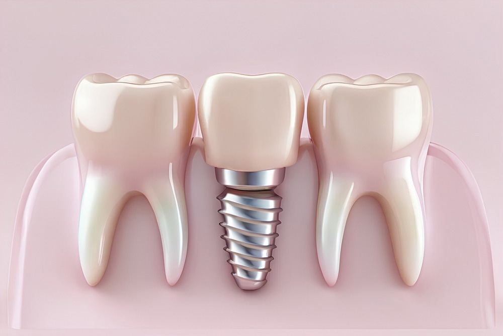
A recent article published in Scientific Reports assessed the cytotoxicity and antibacterial susceptibility of two different ratios of pectin-chitosan polyelectrolyte composite (PCPC) by coating them over commercially pure titanium (CpTi) substrates using electrospraying. The developed PCPC was analyzed for potential application in dental implants.

Image Credit: DentalMan/Shutterstock.com
Background
Dental implants are widely used to fabricate fixed, detachable, and maxillofacial prostheses. These are primarily made from titanium or titanium alloys because of their exceptional biocompatibility, mechanical properties, and corrosion resistance. However, further improvement is required to stabilize the implant and enhance its integration at the bone-biomaterial interface.
Biopolymers such as chitosan (a polysaccharide derived from chitin) and pectin (a plant-based polysaccharide) are promising coating materials to enhance the surface properties of dental implants. While chitosan prevents the creation of a fibrous tissue capsule around medical implants, pectin, with a composition similar to the extracellular matrix of mammals, facilitates cellular adhesion.
This study designed a nanocoating for dental implants based on natural (chitosan and pectin) and synthetic polymers (polyvinyl alcohol (PVA)). PVA, a cost-effective artificial polymer, offers non-toxicity, biocompatibility, and biodegradability, relevant to biomaterial and biomedical applications.
Methods
PCPC solutions were prepared using chitosan (from shrimp shells), pectin (from citrus peel), and PVA with two ratios of pectin-chitosan, 1:2 and 1:3. CpTi grade II discs (diameter: 18, 10, and 6 mm; thickness: 2 mm) were used as substrates for coating with PCPC.
The electrospinning/spraying coating process was performed in a transparent sealed container at room temperature and 40-60 % relative humidity using a high-voltage generator (20 kV). The resulting PCPC coating on CpTi samples was maintained for 24 hours under ambient conditions to evaporate residual acid and water.
The MTT (3-(4,5-dimethylthiazol-2-yl)-2,5-diphenyltetrazolium bromide) assay was employed to measure cell proliferation and cytotoxic effects substrates coated with PCPC (1:2) and PCPC (1:3) and compared to the uncoated CpTi substrates. The evaluation was performed on isolated fibroblast-like cells.
Inhibition zone diameter (IZD) was calculated to examine the antibacterial activity of uncoated and PCPC-coated CpTi substrates via the Kirby-Bauer disk diffusion method. Total anaerobic bacteria extracted from the depth of peri-implantitis were the model microorganisms in these experiments.
The adhesive strength of PCPC coatings was analyzed through pull-off adhesion tests. Their morphology and surface characteristics were studied using atomic force microscopy (AFM) and field-emission scanning electron microscopy (FESEM). In addition, energy dispersive X-ray spectroscopy (EDS) was performed to determine the elemental composition of materials. Fourier-transform infrared (FTIR) spectroscopy ascertained their surface chemical composition.
Results and Discussion
The cytotoxicity test conducted on human fibroblast-like cells after different exposure durations on uncoated, PCPC (1:2)-coated, and PCPC (1:3)-coated CpTi substrates exhibited varying mean cell viability. It generally decreased with time for all substrates, indicating the materials’ biocompatibility and non-cytotoxicity.
PCPC (1:3) exhibited the maximum mean cell viability of 93.42, 89.88, and 86.85 % after 24, 48, and 72 hours, respectively. This coating exhibited maximum IZD against anaerobic bacteria in the Kirby-Bauer disk diffusion test for antibacterial susceptibility. However, the IZD for PCPC (1:3) and (1:2) decreased over time, indicating a decreasing antibacterial effect.
The mean pull-off adhesion strength was 521.6 psi for PCPC (1:3), substantially higher than 419.5 psi for PCPC (1:2). Additionally, the former exhibited better nano-roughness parameters in AFM than other substrates.
FESEM images revealed the uniform surface of CpTi, which had numerous peaks and valleys but no visible defects or fractures. The PCPC (1:2) coating was uniformly deposited on CpTi substrates, with nano-sized structures interspersed with small and large spherical particles. Additionally, the coating layer was devoid of fractures.
Alternatively, FESEM images of PCPC (1:3)-coated CpTi substrates revealed a consistent coating with a closely interwoven network configuration. This coating comprised nano-sized fibers and spherical nanoparticles of various sizes, strongly held together and free of cracks.
The surface features of the PCPC (1:3) coating were more uniform than those of the PCPC (1:2) coating, indicating a more cohesive structure. Finally, FTIR measurements revealed that both PCPC coatings exhibited a purely physical blending in the composite coating without any chemical reaction.
Conclusion and Future Prospects
The researchers successfully demonstrated the potential application of PCPC nanocoatings comprising natural and synthetic polymers in dental implants. The CpTi substrates coated with PCPC (1:3) exhibited enhanced biocompatibility, antibacterial effects, surface nano-roughness, and adhesion strength. Thus, it was considered a highly efficient coating for titanium dental implants.
However, in-vivo studies are important to validate the results of in-vitro studies presented in this study. The researchers suggest conducting animal and clinical studies to validate the proposed coatings' safety, efficacy, and biocompatibility. This will ensure the reproducibility and scalability of the coating composition and deposition method.
Successful implantation of PCPC coatings in clinical settings could transform dental treatments, improving therapeutic outcomes and reducing the risks of implant-related complications.
Journal Reference
Alsharbaty, MHM., Naji, GA., Ghani, BA., Schagerl, M., Khalil, MA., Ali, SS. (2024). Cytotoxicity and antibacterial susceptibility assessment of a newly developed pectin-chitosan polyelectrolyte composite for dental implants. Scientific Reports. DOI: 10.1038/s41598-024-68020-
Disclaimer: The views expressed here are those of the author expressed in their private capacity and do not necessarily represent the views of AZoM.com Limited T/A AZoNetwork the owner and operator of this website. This disclaimer forms part of the Terms and conditions of use of this website.