 By Muhammad OsamaReviewed by Lexie CornerMay 27 2025
A recent study published in Advanced Materials introduces a new approach in 3D bioprinting using a cysteine-modified hyaluronic acid (HA-Cys) bioink.
This formulation is dynamically crosslinked through disulfide bonds facilitated by potassium iodide (KI), improving gelation time, mechanical stability, and biocompatibility.
The goal was to support the fabrication of complex, cell-laden structures for applications such as in vitro disease models, including osteoarthritis, in tissue engineering and regenerative medicine.

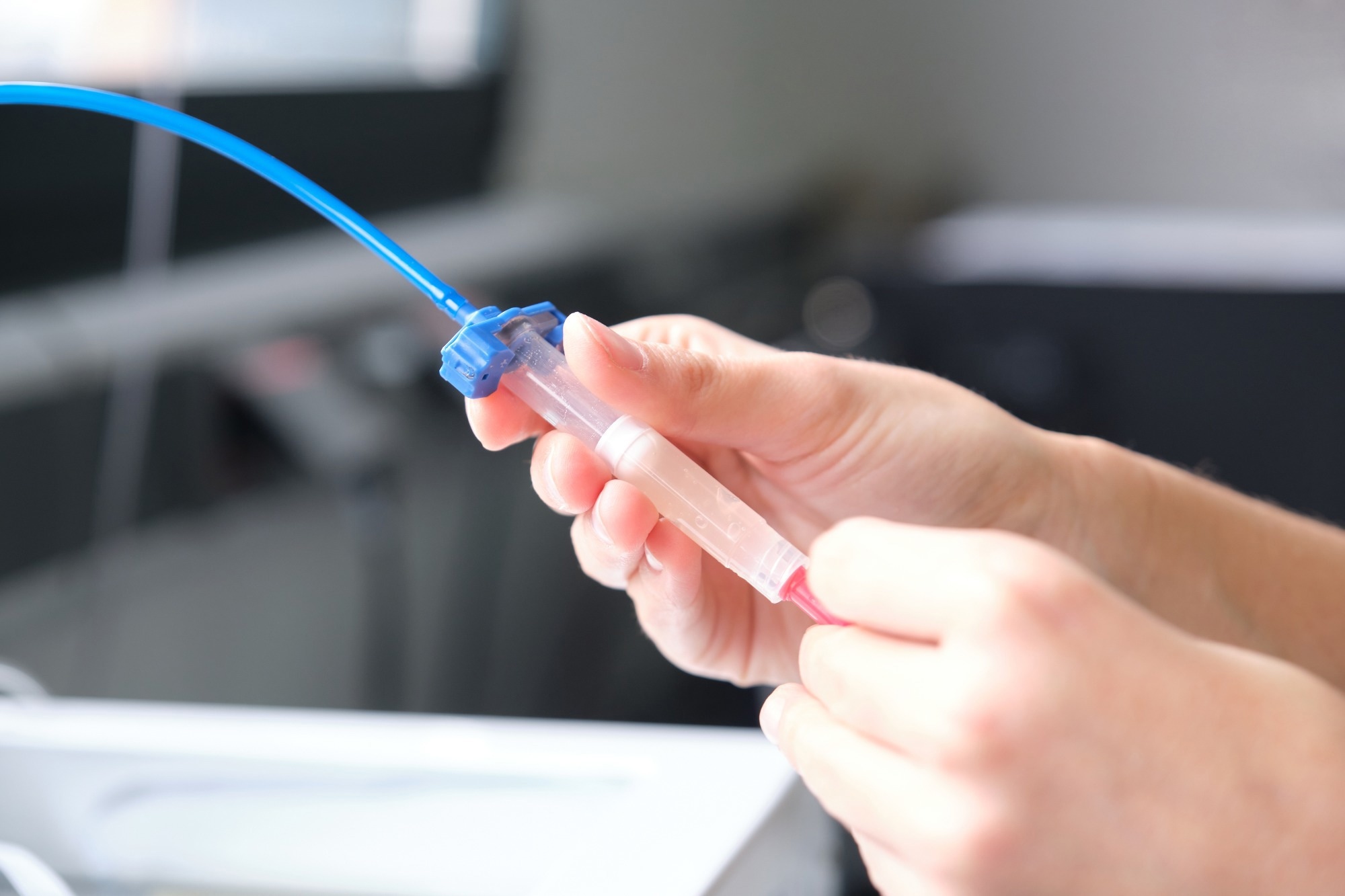
Image Credit: Ladanifer/Shutterstock.com
Background: Challenges in 3D Bioprinting
3D bioprinting enables the construction of biological structures by depositing bioinks—combinations of biomaterials and living cells—in a layer-by-layer process. A key challenge is developing bioinks that balance biocompatibility with mechanical integrity and rapid, stable gelation.
Hyaluronic acid (HA), a naturally occurring polysaccharide in the extracellular matrix, is commonly used due to its favorable biological properties. However, conventional HA-based hydrogels typically exhibit slow gelation and limited mechanical strength.
Tailoring Hydrogel Properties for Precision Bioprinting
This study presents the development of an HA-Cys bioink designed to address limitations in gelation rate and mechanical stability commonly associated with traditional disulfide-crosslinked hydrogels. HA was modified with cysteine groups using carbodiimide coupling chemistry, enabling disulfide bond formation under physiological conditions.
KI was incorporated to improve gelation kinetics and reduce oxidative stress, functioning both as a crosslinking agent and a radical scavenger. The bioink was tested for suitability in extrusion-based bioprinting using ultra-fine nozzles (30G, 108 µm), with attention to maintaining structural coherence and cell viability during printing.
The researchers evaluated the material’s mechanical and rheological properties across different KI concentrations, including storage and loss moduli, gelation time, and viscoelastic behavior. Biocompatibility was assessed through live/dead staining to measure cell viability.
Additionally, the bioink’s ability to maintain cellular activity was investigated by encapsulating human mesenchymal stromal cells (hMSCs) and chondrocytes, and monitoring their metabolic function and proliferation following bioprinting.
Key Findings: Hydrogel Performance and Cellular Viability
The HA-Cys bioink preserved high cell viability (>95 %) and enabled interactions between different cell types, essential for modeling tissue dynamics and disease mechanisms.
Incorporating 50 mM KI reduced gelation time from over an hour to approximately 11.7 minutes, allowing a practical printing window of more than three hours. This rapid gelation supported smooth extrusion through ultra-fine needles without clogging and facilitated the fabrication of complex 3D structures over 3 cm in size with high shape fidelity (up to 90 %).
Rheological assessments confirmed that the bioink exhibited shear-thinning and self-healing properties, essential for maintaining structural integrity post-printing. Mechanical testing indicated that lower KI concentrations did not compromise hydrogel stiffness, while higher levels led to reduced strength due to overly rapid crosslinking.
The antioxidant properties of KI enhanced the hydrogel's radical scavenging activity, achieving a maximum efficiency of 88.8 % at 50 mM. This feature reduces oxidative stress during printing, protecting sensitive cells such as hMSCs.
Biocompatibility studies demonstrated that the hydrogel supported the survival and function of encapsulated hMSCs and chondrocytes over time, successfully creating an osteoarthritis disease model and offering insights into potential therapeutic strategies.
Applications in Biomedical Research
The HA-Cys bioink was evaluated for its potential use in tissue engineering and regenerative medicine, particularly for constructing cell-laden 3D structures intended for in vitro disease modeling. Its formulation supports the inclusion of multiple cell types and can be adapted to mimic features of native extracellular matrix (ECM).
The use of dynamic disulfide crosslinking allows for tunable degradation rates, which may enable controlled release of encapsulated cells or bioactive molecules under specific conditions.
The material's combination of mechanical stability, gelation behavior, and biocompatibility makes it applicable to areas beyond disease modeling, including drug testing, toxicity screening, and regenerative applications. Its capacity to support co-culture systems can help enhance the biological relevance of experimental models, potentially contributing to more representative preclinical data.
Download your PDF copy now!
Conclusion and Future Work
This study presents an HA-Cys-based hydrogel system that incorporates disulfide crosslinking and potassium iodide to improve gelation kinetics and reduce oxidative stress.
The resulting bioink addresses common limitations of HA-based materials, offering a platform suitable for constructing cell-laden hydrogels with preserved cell viability and structural integrity.
Further research could explore additional formulation adjustments for specific clinical or experimental needs, the integration of multi-cellular arrangements, and the use of computational tools to optimize printability and biological performance.
These efforts may support broader applications in tissue engineering, disease modeling, and related biomedical fields.
Journal Reference
Tavakoli, S., et al. (2025). Ultra-Fine 3D Bioprinting of Dynamic Hyaluronic Acid Hydrogel for in Vitro Modeling. Advanced Materials. DOI: 10.1002/adma.202500315, https://advanced.onlinelibrary.wiley.com/doi/10.1002/adma.202500315
Disclaimer: The views expressed here are those of the author expressed in their private capacity and do not necessarily represent the views of AZoM.com Limited T/A AZoNetwork the owner and operator of this website. This disclaimer forms part of the Terms and conditions of use of this website.