This article focuses on atomic force microscopy, electrochemical impedance spectra, and the interrelation between the two concepts. Image processing of anchored bilayers, such as tethered bilayer membranes (tBLMs), using atomic force microscopy (AFM), can identify the type of barrier harm caused by pore-forming enzymes and anticipate the electrochemical impedance spectroscopy (EIS) behavior of such materials.

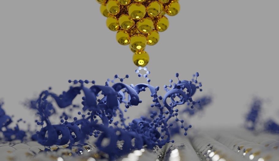
3D rendering of a surface showing the tip of atomic force microscopy (AFM) and protein structures. Image Credit: sanjaya viraj bandara/Shutterstock.com
What is Atomic Force Microscopy?
Atomic force microscopy (AFM) is a popular surface examination method for micro/nanostructured coverings. This adaptable technology may be used to acquire high-resolution nanoscale pictures and investigate local areas in the presence of air (standard AFM) or solution (electrochemical AFM). AFM is increasingly being utilized to explore the interactions of lipid bilayers with proteins such as pore-forming toxins (PFTs) and barrier-disruptive peptides.
AFM can detect peptide implantation, variable protein dispersion in walls in phase-segregated surfaces, development of rings of PFTs, and other structural features critical for understanding how barrier proteins interact with the cellular membrane.
Utilization of Atomic Force Microscopy
AFM is an excellent tool for characterizing nanoparticles and nanomaterials. It provides both descriptive and analytical data on a variety of physical attributes such as size, shape, surface characteristics, and smoothness. Many researchers from all around the globe employ atomic force microscopy in material science applications.
Nanosized chemical, biomechanical (flexural strength, flexibility, viscous, resistive), electric, and electromagnetic characteristics may be obtained via AFM. In comparison to other microscopy techniques, AFM has a lower cost, is easier to use, and can image at atomic resolution. Statistical data, such as dimension, size distribution, and volume ratios, can also be obtained.
A wide range of particle sizes, from 1 nm to 8 nm, may be described in the single scan. AFM provides great resolution and visibility in 3D pictures from tip mobility. AFM is increasingly being utilized to explore the interactions of lipid bilayers with proteins such as pore-forming toxins (PFTs) and barrier-disruptive peptides.
The AFM can scan the morphology of delicate biological matter in their natural settings. It may also be used to investigate the mechanical characteristics of cells and extracellular matrices, such as inherent tensile properties and receptor-ligand associations.
Limitations of Atomic Force Microscopy
Although AMF has been utilized by various industries all over the world for many different purposes, several limitations still are a hurdle in its vast commercialization. The line scan picture size of AFM is a limitation when compared to the scanning electron microscope (SEM). It can only scan a single 150 x150nm nanosized picture at a time.
AFM's scanning speed is also a constraint. Traditionally, AFM cannot scan pictures as quickly as an SEM, with a typical scan taking several minutes. Because of the comparatively slow pace of scanning during AFM imaging, thermodynamic instability in the picture frequently occurs, making the AFM microscope less suitable for determining exact measurements between geographical features on the imaging.
Image artifacts, as with any other imaging technology, are possible. They might be caused by an incompatible tip, a bad working condition, or even by the material itself. Along with this, it also has a restricted vertical range and resolution.
Introduction to Electrochemical Impedance Spectroscopy
For thorough investigations of the electrical effects of PFTs in surfaces, electrochemical impedance spectroscopy (EIS) is the preferred method.
More from AZoM: Exploring Thin Films and Coatings with AFM
The EIS allows for access to the dielectric characteristics and conductivity characteristics of tBLMs and, in certain circumstances, offers insight into the lateral dispersion of flaws in barriers while not being a design pattern per se.
It is a material science analytical tool that may be used to examine mass transfer, chemical reaction mechanisms, dissolution, dielectric characteristics, flaws, crystal structures, and permeability in solids. It is also used to evaluate the effectiveness of chemical sensing and fuel cells, as well as in electrochemical reactions for the research of live cellular membrane activity.
Interrelation Between AFM and EIS
Both AFM and EIS are employed in tandem to describe the form and composition of PFTs in barriers. Although the practical abilities to perform both procedures on the same test specimens are basic, no attempts have been made to statistically connect morphological information recorded by AFM with transmembrane conductivity measurements performed by EIS.
Such comparison assessments would be extremely useful for both single and multiple membrane assemblies. Substantial improvements have recently been achieved in the advancement of EIS data processing. Theoretical research revealed that the number of regenerated protein particles per surface area may be calculated from EIS spectral information. Nonetheless, such theoretical ideas need to be rigorously tested using evidence from alternative structural techniques like AFM.
Research Findings
Researchers from Lithuania, in a recent study, have investigated the feasibility of predicting the electrochemical impedance spectra of membranes containing reconstituted PFTs using AFM pictures. The potential benefit of the innovative technique over subjective defect identification was proven by using a convolutional neural network for the specified entity segmentation task.
Utilizing three different samples of tBLMs, it was discovered that, although non-identical, actual and AI-derived groups of fault dimensions are produced by FEA simulating comparable EIS curves. The projected placement of the phase minimum in Bode plots of admittance, one of the primary EIS spectral characteristics, was within 2–6 percent of the genuine values.
The scientists determined that an autonomous AI-based AFM image analysis system could anticipate EIS spectra, which can be used to examine crucial material characteristics of tBLMs such as submembrane-specific resistance. Using three different samples of tBLMs, it was discovered that the submembrane resistance is 104.25 ± 0.10 Ω.cm, which is somewhat lower than the previously used value (104.5 Ω.cm). These parameters enable the calibration of tBLM biosensors for the quantitative detection of pore-forming toxin activity.
To conclude, confirmation of the usability of AFM to analyze the shape and concentration of detrimental membrane abnormalities such as pore-forming toxins in tBLMs was presented. This data may be utilized to hypothetically anticipate the EIS performance of tBLMs as well as validate this performance for biosensing.
Further Reading
Raila, T., Penkauskas, T., Ambrulevičius, F. et al. 2022. AI-based atomic force microscopy image analysis allows to predict electrochemical impedance spectra of defects in tethered bilayer membranes. Scientific Reports. 12. 1127. Available at: https://www.nature.com/articles/s41598-022-04853-4
Raila, Tomas, et al. 2022. Electrochemical impedance of randomly distributed defects in tethered phospholipid bilayers: Finite element analysis. Electrochimica Acta. 299. 863-874. Available at: https://www.sciencedirect.com/science/article/pii/S0013468618328676?via%3Dihub
DuToit, Marielle, Edgard Ngaboyamahina, and Mark Wiesner. 2021. Pairing electrochemical impedance spectroscopy with conducting membranes for the in-situ characterization of membrane fouling. Journal of Membrane Science. 618. 118680. Available at: https://www.sciencedirect.com/science/article/pii/S0376738820312564?via%3Dihub
Disclaimer: The views expressed here are those of the author expressed in their private capacity and do not necessarily represent the views of AZoM.com Limited T/A AZoNetwork the owner and operator of this website. This disclaimer forms part of the Terms and conditions of use of this website.