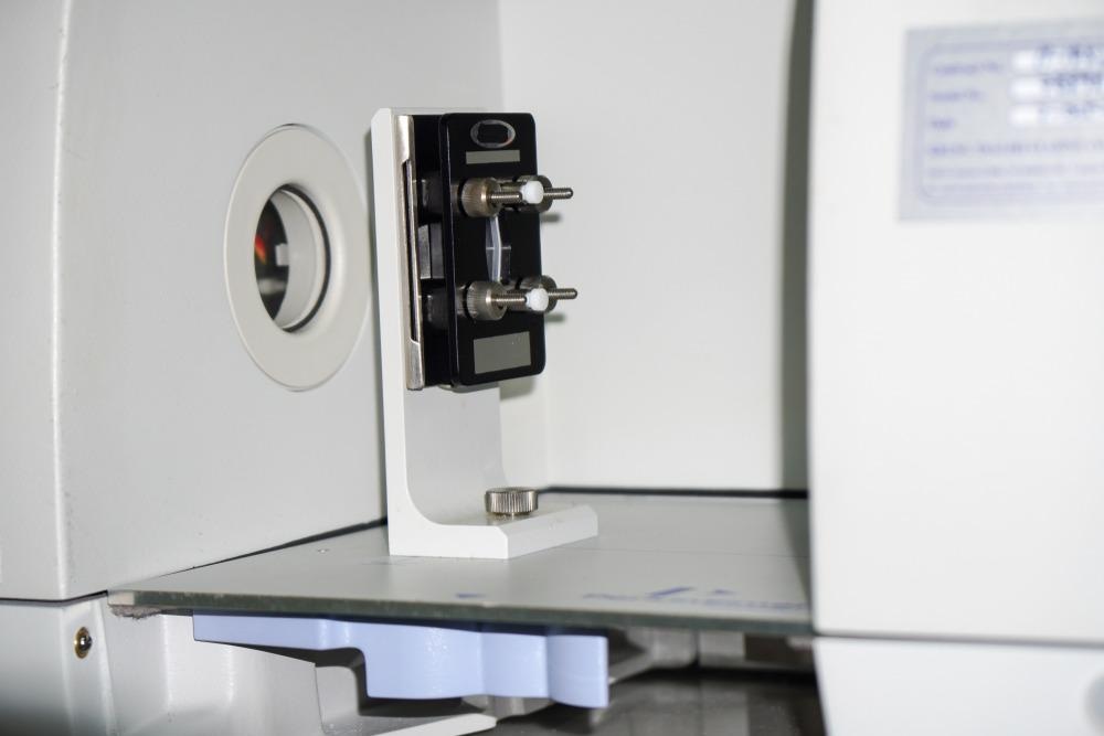
The sample preparation before conducting an infrared spectroscopy (IR) study is as critical as the study itself, and the samples which are difficult to dissolve in any IR-transparent solvent are mixed with potassium bromide (KBr). This article takes a look at the role of potassium bromide in infrared spectroscopy.

Image Credit: boonchok/Shutterstock.com
The electromagnetic (EM) spectrum consists of seven different EM radiations with wavelengths ranging from 10-12 to 103 m. Each radiation type falls within a wavelength bracket that serves as a form of energy in scientific studies. Designing a spectrometer involves a source for specific EM radiation absorbed by the sample and represented as a graph plotted for absorption against wavelength.
IR spectroscopy measures the IR radiation absorbed by a chemical molecule of interest. Each chemical molecule has a set of functional groups which absorb the IR radiation. Irrespective of the structure of the chemical compound, the functional groups tend to absorb IR radiation of the same frequency. The correlation between the molecular structure and the frequency at which it absorbs the IR radiation allows the structural prediction of an unknown molecule.
Practically, low energy mid-IR spectroscopy is preferable over high energy near-IR (NIR) spectroscopy, since the spectra of mid-IR spectroscopy arise from simple vibrational modes of a molecule resulting in cleaner spectra.
Role of Potassium Bromide (KBr) in IR Spectroscopy
The sample preparation before conducting an IR study is as critical as the study itself. Further, the carrier used in preparing the sample should be optically transparent in the IR region (4000-400 cm-1). In this context, alkali halides like potassium bromide (KBr) serve as a carrier with 100% transmittance. The major problem in mid-IR spectroscopy is the high absorbance of radiation by the sample, and this can be resolved by diluting the sample with spectroscopy grade KBr.
The samples which are difficult to dissolve in any IR-transparent solvent are mixed with KBr in IR spectroscopy. The target sample to be analyzed is ground with KBr powder and pressed into a disc, also known as a pellet. The particle size of KBr plays a vital role in pellet preparation.
Sample Preparation with KBr in IR Spectroscopy
If the test sample is amorphous in nature, it is dissolved in a non-aqueous volatile solvent and then deposited as a thin sample layer on the KBr cell by evaporating the solvent.
For the KBr pellet method, it is essential to pulverize the KBr powder into a 200-mesh size followed by drying at 110°Celsius to remove any bound water molecule. For a sample pellet of 13 mm diameter, 0.1 to 1% of the sample is mixed with 200 to 250 mg of powdered KBr. Next, the sample pellet is pulverized and placed into a pellet-forming die. Further, a force of 8 tons is applied under vacuum for several minutes to form a transparent sample pellet, and degassing removes the air and bound water molecules.
In the diffuse reflection method, KBr is packed in the diffuse reflectance accessory sample plate for background measurement, followed by dilution of sample powder in KBr powder and packing the resultant mixture into the sample plate for IR spectrum measurement.
In all the above methods, it is essential to keep the sample and KBr pellet moisture-free since the presence of water molecules gives a broad water peak at 3200 cm-1, which overlaps with functional group peaks (amines, carboxylic acid, alcohol) in the range of 3500-3000 cm-1.
IR Spectroscopy Reveals Protein Structure in KBr Pellets – Research Studies
Fourier transform IR (FTIR) spectroscopy is employed to determine the secondary protein structure in different physical states, including solutions, suspensions, gels, and solids. The most frequent method used in protein structure determination is preparing a KBr pellet of lyophilized protein.
In a study published in the Journal of Pharmaceutical Sciences, the authors determined the structure of two proteins, lysozyme, and α-chymotrypsinogen, for characteristic amide bond determination using FTIR. The results revealed that KBr processing is a convenient and viable option for determining the structure of dried proteins and formulation.
In another study published in the journal Analytical Biochemistry, the authors analyzed the secondary structure of 13 globular proteins in the KBr pellet through FTIR. The results showed a better correlation for α-helix and β-helix in the band of amide I, where the protein secondary structure was conserved in a solid-state and packed into a KBr pellet. The singular value decomposition (SVD) analysis showed that the absorbance of proteins in the KBr pellet was accurate compared to their absorbance in solution. Further, the authors found that the protein secondary structure in the solid-state is a better way to study water-insoluble proteins, fibers, and cereal reserve proteins than their dissolution in organic solvents.
In another study published in Talanta, the authors devised a chemometric method to correct the spectra of materials in the KBr disk by eliminating the water interference. It is a common problem in IR analysis of proteins and polysaccharides using the KBr disk technique. The resulting protein spectra were much more accurate than attenuated total reflection (ATR) spectra.
Conclusion
To summarize, KBr is optically transparent in the fingerprint region of IR spectroscopy and hence is used as a carrier in the form of a disk or pellet. Structural determination of sensitive biomolecules like proteins was possible by employing FTIR and the KBr disk technique, especially in determining their primary and secondary structures.
More from AZoM: What Multi-Analytical Techniques are Used to Assess Paintings?
References and Further Reading
Ingebrigtson, D. N., and Smith, A. L. (1954). Infrared analysis of solids by potassium bromide pellet technique. Analytical Chemistry, 26(11), 1765-1768. https://pubs.acs.org/doi/10.1021/ac60095a023
Forato, L. A., Bernardes-Filho, R., and Colnago, L. A. (1998). Protein structure in KBr pellets by infrared spectroscopy. Analytical Biochemistry, 259(1), 136-141. https://pubmed.ncbi.nlm.nih.gov/9606154/
Ng, L. M., and Simmons, R. (1999). Infrared spectroscopy. Analytical Chemistry, 71(12), 343-350. https://pubs.acs.org/doi/abs/10.1021/a1999908r
Meyer, J. D., Manning, M. C., and Carpenter, J. F. (2004). Effects of potassium bromide disk formation on the infrared spectra of dried model proteins. Journal of Pharmaceutical Sciences, 93(2), 496-506. https://pubmed.ncbi.nlm.nih.gov/14705205/
Gordon, S. H., Harry-O'kuru, R. E., and Mohamed, A. A. (2017). Elimination of interference from water in KBr disk FT-IR spectra of solid biomaterials by chemometrics solved with kinetic modeling. Talanta, 174, 587-598 https://www.sciencedirect.com/science/article/pii/S0039914017306756
Disclaimer: The views expressed here are those of the author expressed in their private capacity and do not necessarily represent the views of AZoM.com Limited T/A AZoNetwork the owner and operator of this website. This disclaimer forms part of the Terms and conditions of use of this website.