X-ray analytical instrument manufacturer Rigaku Corporation is pleased to announce its attendance at the 54th annual meeting of the Texas Society for Microscopy (TSM). The event is hosted by the Kleberg Advanced Microscopy Center at The University of Texas in San Antonio and takes place February 21st - 23rd, 2019. Several workshops are scheduled, as well as a scientific program with platform and poster presentations, along with a vendor exhibition.
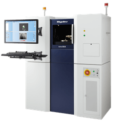
 The Rigaku nano3DX X-ray microscope
On Friday February 22nd, Aya Takase from Rigaku will present “High Resolution X-Ray Computed Tomography for Foam Characterization” – a study, in which a high-resolution, high-contrast and high-speed laboratory X-ray microscope with true submicron resolution was used to characterize foam materials. X-ray microscopes can visualize the solid walls and cellular structure in foam materials in 3D without having to slice and potentially alter the cell structure, enabling the study of cell morphology in detail and calculation of parameters such as porosity and cell volume distribution.
Rigaku, a global leader in X-ray analytical technology, is presenting its latest X-ray microscopy (XRM) and computed tomography (CT) solutions at the meeting. XRM and CT equipment from Rigaku enable nondestructive analysis of large samples at high resolution.
The Rigaku nano3DX X-ray microscope features parallel beam geometry that makes true submicron 3D imaging possible. The Rigaku MicroMax-007 HF rotating anode X-ray generator effects characteristic X-ray radiation for high contrast and rapid data collection, and the ability to easily switch anode materials to optimize contrast for specific sample types.
The nano3DX X-ray microscope is a well-suited X-ray CT scanner for low-density materials such as food, pharmaceuticals, plants, insects, foam and various composite materials.
Computed tomography reveals, at high-speed, the high-resolution, three dimensional structure of an object by means of computer-processed combinations of numerous X-ray images taken from different angles. CT solutions from Rigaku include the Rigaku CT Lab GX industrial 3D X-ray micro CT imager, an ultra-high-speed, high-resolution 3D CT system suited for measurements of pharmaceuticals, medical devices, bones, ores, electronic devices, batteries, aluminum castings, and printed circuit boards.
The CT Lab GX series is designed for ease-of-use, featuring the latest 3D CT technology for measurement of industrial products in a short period of time. The unit’s Sample-Stationary scanning method allows quick and easy sample mounting.
A principal feature of the CT Lab GX imager is its capacity for ultra-high-speed measurement. A CT scan can be achieved in 8 seconds and image reconstruction in 10 seconds. High-definition 3D observation is possible with a minimum resolution of 4.5μm.
Two versions are available: the low-energy “CT Lab GX90,” suited for measurement of subjects such as small animals and low-density materials, and the high-energy “CT Lab GX130,” suited for subjects less penetrable by X-ray beams such as ceramics and metals.
The Rigaku CT Lab HX high-performance benchtop X-ray micro CT system features a combination of the high power X-ray source and the largest field of view (FOV) in its class. Built for easy operation, the compact yet powerful micro CT system offers a large FOV of up to 200 mmφ, and can provide three dimensional X-ray images of a wide variety of samples. Preset scans, programed for typical conditions, require only a few steps to run. The high-power 130 kV, 39 W X-ray source facilitates high-speed image acquisition as fast as 18 seconds/scan.
More information about X-ray imaging solutions from Rigaku is available at www.rigaku.com/products/imaging