May 15 2019
At the University of Bonn, researchers have come up with a novel technique that makes it possible to photograph enzymes at work.
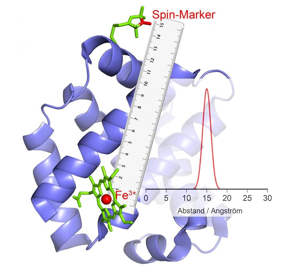
 If it changes its polarity, this causes an echo in the magnetic marker, from which the distance can be calculated. (Image credit: AG Schiemann/Uni Bonn)
This latest technique could provide more insights into the functions of major biomolecules. In addition, the researchers hope to gain a better understanding of the causes of specific enzyme disorders. The new study will soon be featured in the journal, “Chemistry - A European Journal” but it is already available online.
If an extraterrestrial comes across an image of a pair of scissors for the very first time in a craft supplies catalog, he might not have any idea as to what that thing is used for. However, if he is shown a video in which a pair of scissors close and open, then perhaps he might be able to understand their function with some amount of imagination.
Researchers adopt a very similar method when they want to figure out the way an enzyme works—if at all they know the molecular structure, then usually it will only be a still image. They are clueless as to how the enzymes work in action, which parts move towards one another, and which parts move away from one another.
Certain chemical reactions in the cells are catalyzed by enzymes, similar to paper-cutting scissors. The enzymes possess catalytic centers (the blades), which make contact with the starting material, that is, the paper.
The three-dimensional form of the enzyme usually changes during this process. Normally, these conformational changes cannot be made visible, or only with great effort. This often makes it difficult to comprehend the catalysis mechanism.
Dr Olav Schiemann, Professor, Institute of Physical and Theoretical Chemistry, University of Bonn
Schiemann’s research team has successfully created a technique through which the movements of the protein parts against one another can be determined in the course of catalysis. For a number of years, the Bonn researchers have been working on such techniques with remarkable success. In their present work, they have assessed a specifically vital group of enzymes. These enzymes are capable of carrying metal ions with many unpaired electrons in their catalytic centers. Hemoglobin is one such example, which has the potential to bind oxygen with the aid of an iron ion and can, therefore, be transported in the bloodstream.
Flipping ions
“Our current methods are unsuitable for such high-spin ions,” clarified Schliemann’s colleague Dr Dinar Abdullin. “We therefore developed a new method, worked out the theory and successfully tested it.”
The scientists leveraged the fact that high-spin ions act like tiny electromagnets. Moreover, they can arbitrarily alter their polarity, that is, they “flip”—the South Pole becomes the North Pole, and the North Pole becomes the South Pole. A phenomenon like this can be utilized for measuring distance. Here, the researchers connect the enzyme with specific chemical compounds that also possess electromagnetic characteristics.
“When the high-spin ions flip, these small electromagnets react to the changed magnetic field in their environment by also changing their polarity,” explained Dr Abdullin. How and when they do this is dependent, among other things, on the distance to the high-spin ion. As a result, the distance between the two can possibly be determined accurately.
If a single enzyme is bound with a number of magnetic groups, the distance of all these magnetic groups to the high-spin ion and therefore to the catalytic center is achieved.
By combining these values, we can measure the spatial position of this center, as if we were using a molecular GPS. For example, we can determine how its position changes relative to the other magnetic groups in the course of catalysis.
Dr Olav Schiemann, Professor, Institute of Physical and Theoretical Chemistry, University of Bonn
Nevertheless, the researchers are yet to actually watch the enzyme at work.
We are still working with frozen cells. These contain numerous enzymes that were frozen at different points in time during the catalytic reaction. So we do not obtain a film, but a series of “stills”—as if the scissors from the introductory example were photographed at countless different moments during the editing process.
Dr Olav Schiemann, Professor, Institute of Physical and Theoretical Chemistry, University of Bonn
“But we are already working on the next improvement,” emphasized the chemist: “The spatial measurement of biomolecules in cells and at room temperature.”
The team is hoping to get a better understanding of the development of specific diseases that are activated by the enzymes’ functional disorders. Apart from Dr Maxim Yulikov of ETH Zurich from the University of Bonn, the working team headed by Professor Dr Stefan Grimme (also Institute of Physical and Theoretical Chemistry) and Professor Dr Arne Lützen of Kekulé Institute was also involved in the study.