The X-ray microtomography lab at Luleå University of Technology (LTU), Sweden shares some interesting insights on 3D quantitative analysis of snow compaction. The snow sample shows fresh snow obtained only minutes after snowfall. The width of the snow crystal is around 0.85 mm, and the width (diameter) of the center tunnel ranges from 60 micron (at the entrance) to 220 micron on the opposite side.
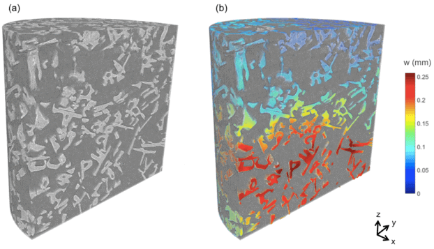
Initially, the sample bed measured 5 mm in height and 6 mm in diameter. A spatial resolution of 4 micron was used to perform scans, and a Deben CT5000TEC load stage with a 500 N load cell was used to carry out in-situ loading. At a constant temperature of -15 °C, four XMT scans were obtained along the load cycle 0N-10N-18N-25N. The software Dragonfly Pro (ORS) was then used to obtain Quantitative analysis of microstructure (shape of crystals, porosity etc.) from 3D image analysis, and finally the compaction of the snow bed was analyzed by digital volume correlation (DVC) using software from LaVision.
Credit: X-ray microtomography lab at Luleå University of Technology (LTU), Sweden
One major advantage of using the Deben stage is the flexibility in using various load cells based on material and application. The easy-to-use software interface is supported by ZEISS’ Scout and Scan software, which is used for controlling ZEISS Xradia Versa.
A recent study that we are very proud of is 3D quantitative in-situ imaging of micro scale snow crystals and how they respond to compaction. This study was quite challenging, and required a lot of careful planning, but turned our very well. It’s received a lot of positive feedback from both the scientific community as well as from media. In fact, the first results from this study were broadcast on national television (in Sweden) only a few hours after performing the scans. These measurements would have been hard to achieve without the Deben CT5000TEC stage as they required precise control of both the mechanical load and the temperature (freezing capability) at the same time.
Fredrik Forsberg, Associate Senior Lecturer, Luleå University of Technology

3D visualization of snow crystals in bed of fresh snow (a). Quantitative analysis of snow compaction in bed when loaded at 10 N (b).
Conclusions: 3D Quantitative Analysis of Snow Compaction
Digital volume correlation and X-ray microtomography enable the LTU researchers to perform quantitative in-situ studies of snow compaction. In addition, these tools enable them to study the compaction process at multiple spatial scales – grain-to-grain interactions and global phenomena.
X-ray microtomography and the use of a load stage with temperature control, for example, Deben CT 5000TEC, enables studies of snow compaction behavior and snow crystal microstructure in a way that had not been possible until now.

This information has been sourced, reviewed and adapted from materials provided by Carl Zeiss Microscopy GmbH.
For more information on this source, please visit Carl Zeiss Microscopy GmbH.