Aristotle’s postulate that “nature abhors a vacuum” could be used to explain why X-ray photoelectron spectroscopy (XPS) has not found routine application in the analysis of biological samples.
The requirement of an XPS instrument to operate at ultra-high vacuum is at odds with the hydrated environments of bacteria and, more generally, the application of biomaterials.

Image Credit: Kratos Analytical, Ltd.
In the late 1980s, Buddy Ratner compared a surface analyst’s perception of a surface with a biologist’s. He stated that ‘on the whole, biologists will not invoke surface-induced effects in their hypotheses. On the other hand, the surface-scientist will consider the problems of biology as being too complex and disorderly to be dealt with using the tools available.1
While the gap between the two disciplines remains, there is evidence to suggest that it is getting smaller. Biologists understand the importance of the surface of biomaterials. Surface analysis of biologically important surfaces is being undertaken and becoming more common.
In their recent publication, Kjaervik et al. compare two different approaches which facilitate XPS analysis of the surface chemistry of a bacterial cell envelope.2
Recent developments in instrumentation allow the analysis of biological samples at near ambient pressure (NAP-XPS) or as frozen-hydrated samples (cryo-XPS).
The cryo-XPS approach relies on a sample fast-freezing technique developed to study processes at the water-mineral interface and subsequently applied to biological samples.2
This sample preparation technique contrasts with the more commonly used freeze-drying approach as the rapid freezing of the hydrated sample retains the amorphous water at the solid surface of the material. Cryo-XPS allows the analysis of intact hydrated microbial cells without damaging or rupturing cell membranes.
The NAP-XPS analysis requires a dedicated spectrometer capable of maintaining the sample at near ambient conditions. For their comparative study, Kjaervik et al. introduced the bacterial sample while dosing a high vapor pressure into the analysis chamber. This ensures that the sample is not exposed to a high or ultra-high vacuum.
Complexity is added to the NAP-XPS analysis due to the attenuation of the photoelectrons at NAP conditions. This is a result of the scattering of electrons by the water vapor in the gas phase.
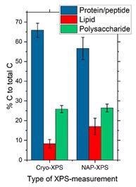
The authors conclude that data from both cryo- and NAP-XPS are in good agreement with each other. Using a method developed by the group at the University of Umeå, the C 1s X-ray photoelectron spectrum was used to identify three spectral components: protein/peptidoglycan, lipid and polysaccharide.
They noted a higher surface content of the lipid-like C at the surface of the bacteria analyzed by NAP-XPS, which they suggest could originate from adventitious carbon contamination.
Importantly, it is proposed that the cryo-XPS method leaves a hydrophilic “protective” hydrated layer, making the sample less prone to surface contamination by aliphatic carbon species.

Figure 1. Bar charts showing the average surface composition of P. fluorescens bacteria sample expressed as the amount of substance related to the different molecular classes constituting the bacterial cell envelope measured under cryo- and NAP conditions from analysis of the respective C1s spectra using the ‘Umeå method.’ Image Credit: reference [2].
This and other recent publications demonstrate that X-ray photoelectron spectroscopy can be used to analyze complex, hydrated samples using either cryo- or near ambient pressure approaches.3,4
Developments in sample preparation, sample handling and modern instrumentation continue to narrow the gap between the disciplines of biologists and surface scientists.
Kratos Analytical manufactures X-ray photoelectron spectrometers used for a wide range of materials characterization. The latest generation AXIS Supra+ spectrometers support cryo-XPS experiments with liquid-nitrogen cooling in the entry and sample analysis chambers.
For more information on XPS analysis of biomaterials using Kratos Analytical XPS instruments, visit https://www.kratos.com/applications/biomaterials
References
- Ratner B.D. ed Surface characterization of biomaterials, Progress in biomedical engineering, vol. 6 Elsevier, 1988
- Kjærvik M, Ramstedt M, Schwibbert K, Dietrich PM and Unger WES (2021) Comparative Study of NAP-XPS and Cryo-XPS for the Investigation of Surface Chemistry of the Bacterial Cell Envelope. Front. Chem. 9:666161. doi: 10.3389/fchem.2021.666161
- C. G. Clarkson, A. Johnson, G. J. Leggett, and M. Geoghegan, Slow polymer diffusion on brush-patterned surfaces in aqueous solution”, Nanoscale 2019, 11, 6052-6061.
- Andrei Shchukarev, Bjorn Sundberg, Ewa Mellerowicz and Per Persson, XPS of a Living Tree, Surf. Interface Anal. 2002; 34: 284–288. DOI: 10.1002/sia.1301

This information has been sourced, reviewed and adapted from materials provided by Kratos Analytical, Ltd.
For more information on this source, please visit Kratos Analytical, Ltd.