Lacustrine turbidites offer a reliable record of seismic activity. A typical sedimentary sequence is produced when a turbidity current moves downslope. If the current generates enough energy, this sequence is comprised of a fine to coarse-grained sand fraction at the bottom with a layer of fine grains of silt or clay toward the top.
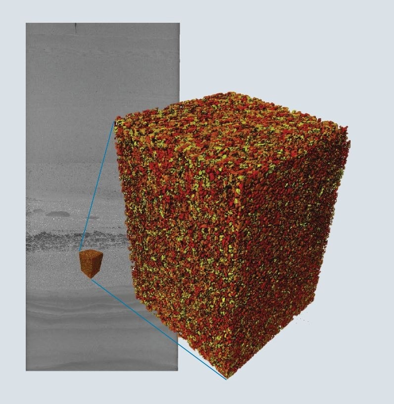
Image Credit: TESCAN USA Inc.
To reconstruct the overall impact of past earthquakes, it is crucial that turbidites are properly characterized at different locations. CT allows for the reconstruction of the orientation and flow direction of sedimentary structures present in core samples.
However, in the absence of sedimentary structures, grain fabric, and size analysis can offer essential information. However, medical CT lacks sufficient resolution for sufficient grain size analysis, which creates a significant demand for non-destructive sediment core analysis to evaluate grain sizes accurately.
Sample
A one-meter-high sediment core sample representing a turbidite section was mounted to the rotational stage of the TESCAN CoreTOM. The sample was extracted from a lake in Alaska to study the paleoseismic activity in the region.
For this sample, the entire section should be evaluated using a high-resolution imaging system.
System
The TESCAN CoreTOM combines the field-of-view of a standard medical-grade CT scanner with high-resolution imaging performance in a single instrument. The built-in workflows are well-suited for multi-resolution imaging going from the core to the grain or pore scale. The core is positioned vertically and then calibrated within the coordinate system of the instrument.
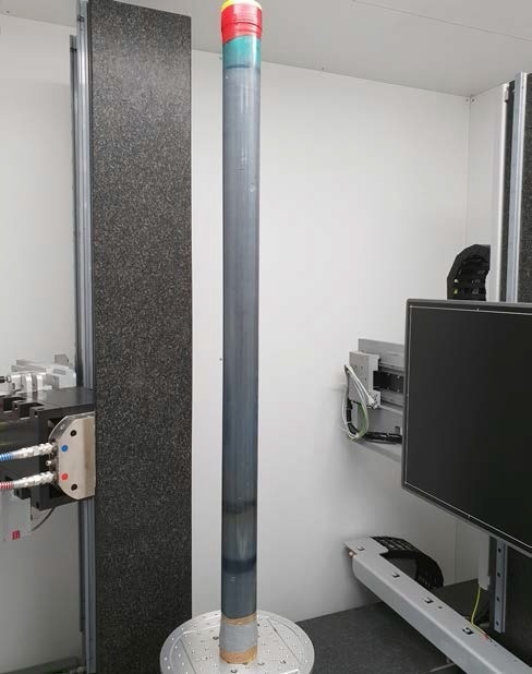
Figure 1. One-meter high sediment core mounted in the TESCAN CoreTOM. Because of the extended vertical travel of the source and detector it is possible to scan complete cores up to 1 m in high resolution. Image Credit: TESCAN USA Inc.
Overview: Image and VOI Selection
Based on the information that a quick overview of the complete core provides, Aquila can be used to prepare a single-acquisition script. The quick overview can be a single radiograph of the complete length or a complete reconstruction. The unparalleled coordinate system allows for the marking of volumes of interest (VOI) directly onto the radiographs or reconstructed volume.
For each VOI, it is possible to store each individual parameter set such as tube, accelerating voltage, exposure, etc. By marking each of the VOIS, a complete script can be constructed that inspects the complete core automatically in line with a multi-scale and multi-resolution process.
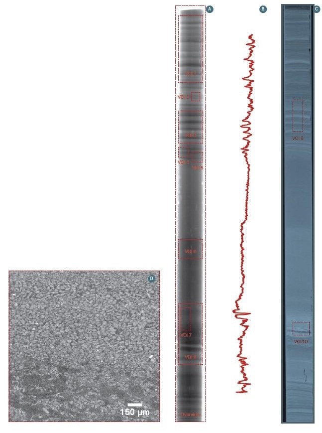
Figure 2. (A) Overview radiograph from the complete core. This can be used to select regions for volume of interest (VOI) scans. (B) Line profile from the acquisition software to fine tune the position of the VOIS. (C) Vertical slice through the reconstructed volume. Also, on this image can be used to select VOIS. (D) Reconstructed slice of a certain VOI scan. Image Credit: TESCAN USA Inc.
Results
For the high resolution VOIS, the grain size distribution can be extracted. This is due to the fact that the coordinates of each grain can be calculated, making it possible to back project their coordinates in the complete overview scan.
Once the grains have been segmented, multiple analyses can be conducted on the volume, including grain size distribution, orientation of the grains in 3D, variation in function of height, etc. The integrated TESCAN CoreTOM coordinate system means that results can later be associated with other regions or even the complete overview scan.
In case of the turbidite section, the fining upward sequence of the grains was analyzed and compared to other regions.
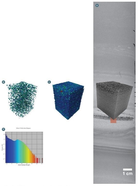
Figure 3. (A) Cross-section through the 40 µm scan with indication of the VOI scanned with 10 µm resolution; (B) Grains showing an orientation with Phi> 90°; (C) Grain size distribution with small grains (in blue), and large grains (in red); (D) Volume/surface area histogram for all the grains. Image Credit: TESCAN USA Inc.
Conclusions
A complete core of 1 m can be scanned in under 10 minutes by a stacked and merged acquisition. This produced a resolution of 80 µm of the entire core. Based on the overview scan, a variety of volume of interests (VOIS) can be defined with a higher resolution acquisition.
Subsequently, all scans are registered in the overview scan by utilizing the integrated coordinate system of the TESCAN CoreTOM. This makes non-destructive grain size analysis of the coarse fraction (> 10 µm) possible.
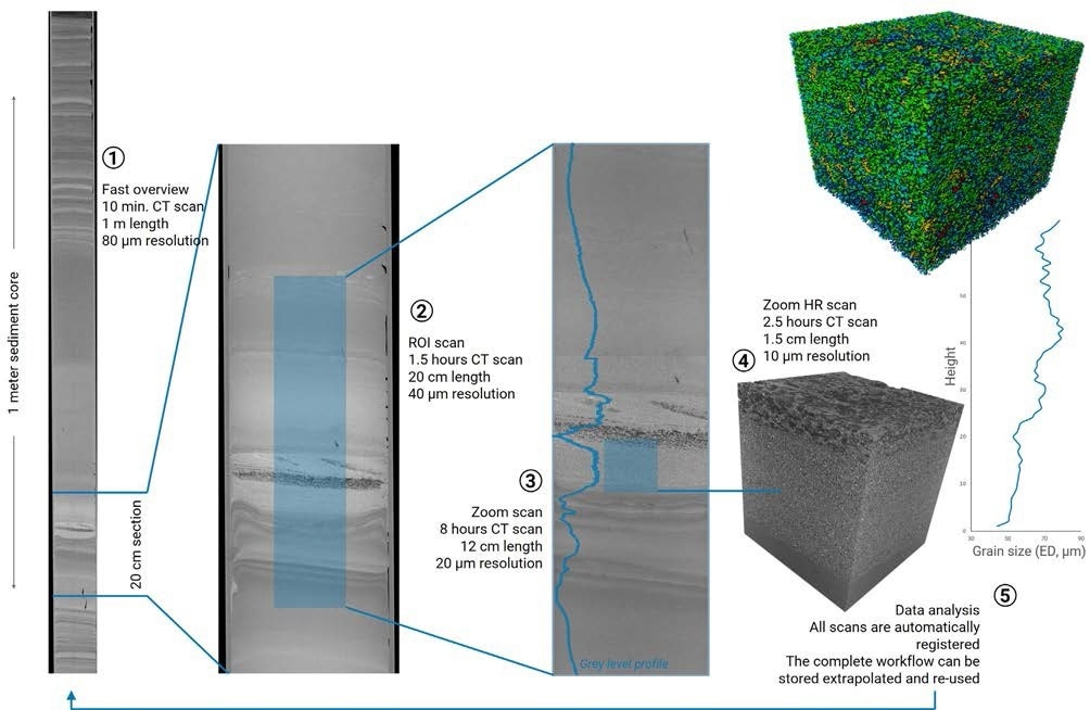
Image Credit: TESCAN USA Inc.

This information has been sourced, reviewed and adapted from materials provided by TESCAN USA Inc.
For more information on this source, please visit TESCAN USA Inc.