Present day oil analysis is done by using optical emission spectroscopy (OES) to measure the ppm (parts per million) levels of wear metals, contaminants and additives in oil samples. This paper provides an overview of Rotating Disc Electrode Elemental Spectroscopy and its use for in-service oil analysis applications.
Today, spectrometric oil analysis finds application in any closed loop lubricating system such as:
- Gas turbines, diesel and gasoline engines
- Transmissions, gearboxes
- Compressors and hydraulic systems.
Table 1 shows typical metal elements can be analyzed by spectroscopy and their sources.
Table 1. Typical source of elements analyzed by spectroscopy in oil
| Metal |
Engine, Transmission, Gears |
Hydraulic Fluid |
Coolants |
| Aluminum Al |
Pistons or Crankcases on Reciprocating Engines, Housings, Bearing Surfaces, Pumps, Thrust Washers |
Pumps, Thrust Washers Radiator Tanks, |
Coolant Elbows, Piping, Thermostat, Spacer Plates |
| Barium Ba |
Synthetic Oil Additive Synthetic Fluid |
Additive |
Not Applicable |
| Boron B |
Coolant leak, Additive |
Coolant Leak, Additive |
pH Buffer, Anticorrosion Inhibitor |
| Calcium Ca |
Detergent Dispersant Additive, Water Contaminant, Airborne Contamination |
Detergent Dispersant additive, Water Contaminant, Airborne Contamination |
Hard Water Scaling Problem |
| Chromium Cr |
Pistons, Cylinder Liners, Exhaust Valves, Coolant Leak from Cr Corrosion Inhibitor |
Shaft, Stainless Steel Alloys |
Corrosion Inhibitor |
| Copper Cu |
Either brass or bronze alloy detected in conjunction with zinc for brass alloys and tin for bronze alloys. Bearings, Bushings, Thrust Plates, Oil Coolers, Oil Additive |
Bushings, Thrust Plates, Oil Coolers |
Radiator, Oil Cooler, Heater Core |
| Iron Fe |
Most common of wear metals. Cylinder Liners, Valve Guides, Rocker arms, Bearings, Crankshaft, Camshaft, Wrist Pins, Housing |
Cylinders, Gears, Rods |
Liners, Water Pump, Cylinder Block, Cylinder Head |
| Lead Pb |
Bearing Metal, Bushings, Seals, Solder, Grease, Leaded Gasoline |
Bushings |
Solder, Oil Cooler, Heater Core |
| Magnesium Mg |
Housings on Aircraft and Marine Systems, Oil Additive |
Additive, Housings |
Cast Alloys |
| Molybdenum Mo |
Piston Rings, Additive, Coolant contamination |
Additive, Coolant Contamination |
Anti-cavitation Inhibitor |
| Nickel Ni |
Alloy from Bearing Metal, Valve Trains, Turbine Blades |
Not Applicable |
Not Applicable |
| Phosphorous P |
Anti-wear Additive |
Anti-wear Additive |
pH Buffer |
| Potassium K |
Coolant Leak, Airborne Contaminant |
Coolant Leak, Airborne Contaminant |
pH Buffer |
| Silicon Si |
Airborne Dusts, Seals, Coolant Leak, Additive |
Airborne Dusts, Seals, Coolant Leak, Additive |
Anti-foaming and Anticorrosion Inhibitor |
| Silver Ag |
Bearing Cages (silver plating), Wrist Pin Bushings on EMD Diesel Engines, Piping with Silver Solder Joints from Oil Coolers |
Silver Solder Joints from Lube Coolers |
Not Applicable |
| Sodium Na |
Coolant Leak, Salt Water and Grease in Marine Equipment, Additive |
Coolant Leak, Salt Water and Grease in Marine Equipment, Additive |
Inhibitor |
| Tin Sn |
Bearing Metal, Piston Rings, Seals, Solder |
Bearing Metal |
Not Applicable |
| Titanium Ti |
Gas Turbine Bearing Hub Wear, Turbine Blades, Compressor Discs |
Not Applicable |
Not Applicable |
| Zinc Zn |
Anti-wear Additive |
Anti-wear Additive |
Wear Metal from Brass Components |
Principles of Spectroscopy
Spectroscopy enables the detection and quantification of elements present in a material. Spectroscopy makes use of the fact that each element has a unique atomic structure and when energy is applied, each element emits light of specific colors or wavelengths.
The principles of spectroscopy include the following:
- It is possible to differentiate the elements as no two elements have the same spectral line pattern.
- The emitted light intensity is proportional to the quantity of the element in the sample enabling the concentration of that element to be determined.
- The light has a particular wavelength or frequency determined by the energy of the electron in transition. As several transitions of different energy is possible for complex atoms that have several electrons light of a number of wavelengths is emitted.
- In case this light is dispersed by using a dispersing element such as a prism, a line spectrum will result. These spectral lines are distinct to the atomic structure of only one element. For the hydrogen atom having atomic number 1, the spectrum is fairly simple as shown in Figure 1.
- The iron spectrum with atomic number 26 is much more complex with many emission lines in the visible spectrum corresponding to the many possible electronic transitions that may occur as shown in Figure 2.
- If more than one element is present in the sample, spectral lines of distinctively different wavelengths will appear for each element. To quantify and identify the elements in the sample, these lines must be separated.

Figure 1. Emission Spectrum of Hydrogen

Figure 2. Emission Spectrum of Iron
Rotating Disc Electrode Optical Emission Spectroscopy (RDEOES)
Spectrometers that view the large number of spectral lines from a heated or an excited sample are called optical emission spectrometers. All optical emission spectrometers consist of three main components, which are:
- Excitation source that introduces energy to the sample
- Optical System, which separates and resolves the resulting emission from that excitation into its component wavelengths
- Readout System that enables the detection and measurement of the light that has been separated into its component wavelengths by the optical system and presents this information to the operator in a usable fashion.
Since early days of spectroscopic oil analysis, oil has been burned or sparked between a rotating carbon disc electrode and a carbon rod electrode. The sample is placed in a sample cap, the disc is partially immersed in the oil sample and the disc rotates as the burn proceeds as shown in Figure 3.
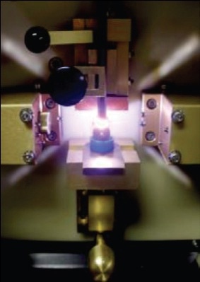
Figure 3. RDE Spectrometer Sample Stand Showing Oil Sample Being "Burned"
This needs about 2 or 3 ml of sample based on the exact cap used. For elimination of sample carryover, a fresh disc and a newly sharpened rod are required. This method is known as the rotating disc electrode (RDE) optical emission spectroscopy (OES) , or combining the two, RDEOES.
An optical system separates the light coming from the plasma into the discrete wavelengths of which it is comprised. An optical device known as a diffraction grating is used to separate the discreet wavelengths.
Figure 4 shows the major components of an oil analysis spectrometer using a polychromator optic based on the Rowland Circle concept.
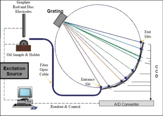
Figure 4. Schematic of a Rotating Disc Electrode Optical Emission Spectrometer for Oil Analysis
During design of a spectrometer the key consideration is the region of the spectrum where the wavelengths of interest occur. Light is emitted by most elements in the visible region of the spectrum. There are elements that emit mainly in the Far Ultra Violet (FUV) region of the spectrum. This is significant as FUV radiation does not transmit well through air; rather, it is mostly absorbed. In order that the optical system is able to view spectral lines, it needs to be mounted in a vacuum chamber or filled with gas transparent to FUV light, so the emitted light can reach the grating, be diffracted, and then be detected at the focal curve. Hence a gas supply system or a vacuum pump and a sealed chamber are part of the system.
A spectrometer’s readout system is controlled by industrial grade software and processor. An amplifier and a clocking circuit reads the charge on a Photo Multiplier Tube or CCD chip and converts it from an analog to digital (ADC) signal to measure the light that has fallen on a pixel. The charge on a pixel is converted to an arbitrary number defined as "intensity" units. Once the analysis is completed, the total intensities for each element are compared to calibration curves stored in memory and converted to the element concentration in the sample.
Concentration is normally expressed in parts per million (ppm). This data can be printed on a printer or displayed on a video screen. On completion of the analysis or recording of results, the system is ready for the next analysis. The analysis results may be left on the screen, stored on the hard disk, or can be sent to an external computer.
A conventional spectrometer of the 1970s is shown in Figure 5. Figure 6 shows the Spectroil Q100, which weighs only 163 lbs (74 kg) with a very small footprint while still maintaining the same analytical capability as the bigger systems in previous generations.
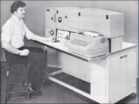
Figure 5. A Direct Reading Oil Analysis Spectrometer from the 1970s
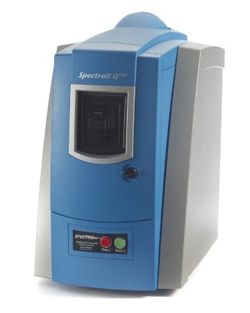
Figure 6. The Spectroil Q100 RDE Spectrometer
Automating the RDE Spectrometer
Challenges for automating the RDE spectrometer are:
- The graphite electrodes need to be replenished after each analysis.
- The rod electrode needs to be sharpened after each burn and also becomes shorter after each sharpening.
The practical solution to RDE spectrometer automation is to use two graphite disc electrodes as shown in Figure 7.
The automation system consists of two parts:
- A robot for exchanging the consumable disk electrodes
- An automatic sample changer - a robotics arm in the sample changer automatically introduces each of 48 oil samples in succession, at a rate of 80 samples per hour and without the need for sample dilution.
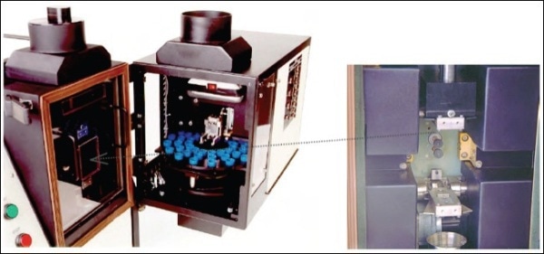
Figure 7. Robotics for RDE spectrometer

Figure 8. Automatic Rotrode Filter Spectroscopy (A-RFS) Fixture
The complete automation system is mounted to the spectrometer sample stand and fulfills all the functions of sequentially introducing and removing oil samples and exchanging graphite electrodes. It is self-contained and works independently of the spectrometer operating software.
Large Particle Size Analysis Capability
One of the shortcomings of spectrometers is the ability to identify and quantify large wear and contaminant particles. Today, the particle size limitation of RDE spectrometers has been eliminated with simple ancillary systems such as the rotrode filter spectroscopy (RFS) method, which involves the following:
- The discs are clamped with a fixture so oil can be drawn through the outer circumference of the discs when a vacuum is applied to the inside of the discs.
- The particles in the oil are captured by the disc.
- The oil is then washed away with solvent, the disc is allowed to dry, and the particles are left on the disc electrode so they are vaporized and detected when run on the RDE spectrometer.
- A multi-station fixture is used so a number of samples can be filtered at one time.
Extended Application Development
The RDE spectrometer is mainly designed for used oil and fuel analysis, several methods and recent enhancements have increased productivity through expanded capabilities.
A used coolant analysis program detects both the coolant condition and the presence of any contaminants or debris. The coolant fluid can be used as a diagnostic medium as the coolant carries heat away from the engine parts as well as fine debris from the interior surfaces of the cooling system. Analyzing the wear debris can offer key data on the condition of the internal parts of the cooling system.
Lubricant mix-ups often occur when an oil system is "topped off' to replace the oil that has been lost due to use or leakage.
Table 2 is a summary of the last four spectrometric oil analyses for a medium speed diesel engine from a locomotive.
Table 2. Spectrometric Results in ppm for an EMD Medium Speed Diesel Locomotive
| |
Fe |
Cu |
Ag |
Mg |
p |
Zn |
| 30-Sep |
19 |
10 |
0 |
0 |
0 |
3 |
| 23-Dec |
21 |
10 |
0 |
0 |
9 |
3 |
| 23-Mar |
27 |
13 |
2 |
107 |
75 |
90 |
| 11-Jun |
25 |
30 |
10 |
220 |
110 |
123 |
Conclusion
In summary, RDE Optical Emission Spectroscopy has gone through a number of changes rendering it suitable for applications in a wide range of industries as a reliable tool to analyze fluid samples for condition monitoring and quality control applications.

This information has been sourced, reviewed and adapted from materials provided by AMETEK Spectro Scientific.
For more information on this source, please visit AMETEK Spectro Scientific.