Sep 9 2016
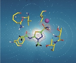
 Neutron crystallography aids in drug design. (Credit: M. P. Blakeley)
Neutron crystallography is a complementary method that is essential for X-ray crystallography as it provides intricate data regarding the hydrogen atom and proton positions in biological molecules.
As neutrons are a non-destructive probe, the resulting structures are free from radiation damage even at room temperature.
Several biological processes, such as enzyme mechanisms can be better understood when knowledge of H-bonding networks, protonation states and water molecule orientations, coupled with details relating to electrostatic and hydrophobic interactions are known. It can also help direct structure-based drug design.
Acetazolamide (AZM), a type of sulphonamide, binds to human carbonic anhydrase isoform II with high affinity. The first-ever neutron crystallography study of a clinically utilized drug adhered to its target was that of AZM. The interconversion of H2O and CO2 to H+ and HCO3- is catalyzed by zinc metalloenzymes such as human carbonic anhydrases (hCA). H+ happens to be a major reaction for fluid secretion, respiration, pH regulation, and many such physiological processes.
hCA isoforms act as major clinical targets for treating many diseases, including epilepsy and glaucoma. One of the 12 catalytically active isoforms is hCA II, and due to sequence conservation between them, significant off-target adherence to other isoforms takes place, which lead to side effects and reduced drug efficiency. Therefore, effective hCA isoform-specific drugs need to be designed.
More than 400 X-ray crystal structures have been established for hCA II, with about 50% of these determined in complex with inhibitors. Despite the availability of X-ray structural data, critical details about the H-atom positions of the solvent and protein, and the bound inhibitor’s charged state were, until recently, missing for all, except for the hCA II/ AZM complex.
In IUCrJ’s September issue, McKenna and the team have described the neutron and X-ray crystallographic studies of hCA II in complex with the methoazolamide (MZM) inhibitor. They provided the missing details of hydrophobic interactions and H-bonding in the complex, and successfully located the MZM’s charged state. The team then compared the AZM-MZM binding in the neutron structures at room temperature and discussed the observed variations in binding in terms of the entropic and enthalpic contributions to drug binding. This indicates that with regard to MZM, hydrophobic forces possibly off-set the loss of a detailed H-bonding network.
In the last few years, an increasing number of neutron structures have been deposited in the Protein Data Bank, which comprises of many other examples of enzyme-drug complexes. Even though the total number of neutron structures is still comparatively small, there are increasing numbers of instances where neutron crystallography has provided the solutions to queries that have remained unanswerable using other methods.
This news article is an excerpt taken from a commentary by Matthew Blakeley.