May 3 2019
It is said that the future of medicine will be biological, and researchers are hoping that 3D-printed biologically functional tissue would soon be used to substitute the body’s permanently damaged tissue.
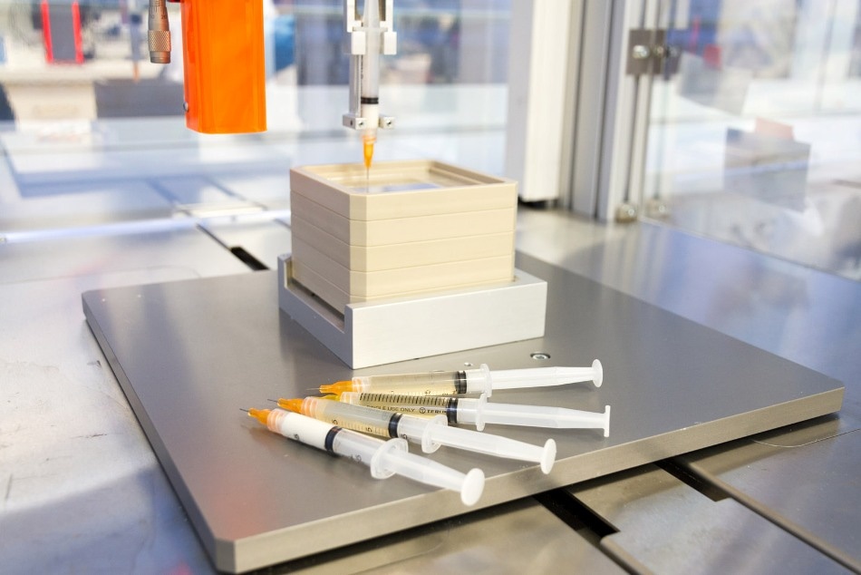
 Syringes containing various bio-ink formulations. (Image credit: Fraunhofer IGB)
Now, a research team at the Fraunhofer Institute for Interfacial Engineering and Biotechnology IGB (Fraunhofer IGB) has been collaborating with the University of Stuttgart for several years on a unique project to create and improve appropriate bio-inks for additive manufacturing. By changing the biomaterial’s composition, the team has already expanded its portfolio successfully to include vascularization and bone inks. That development has laid the basis for producing tissue structures that look like bones and contain capillary networks.
Currently, 3D printing is gaining traction in manufacturing and is also attracting a great deal of interest in the world of regenerative medicine. Now, with the help of this additive manufacturing technique, researchers are hoping to produce bespoke biocompatible tissue scaffolds—scaffolds that will substitute permanently damaged tissues.
At Fraunhofer IGB in Stuttgart, a research team is also working on bio-based inks for making biological implants in the lab through 3D printing methods. In order to develop a 3D object in the preferred pre-programmed shape, the researchers use a layer-by-layer technique to print a liquid mixture containing biopolymers like hyaluronic acid or gelatin, living cells, and an aqueous medium. Such bio-inks continue to be in a viscous state at the time of printing and are subsequently subjected to ultraviolet (UV) light so that they crosslink into water-containing polymer networks known as hydrogels.
Targeted chemical modification of biomolecules
The biomolecules can be chemically altered by researchers to provide the ensuing gels with varying degrees of swellability and crosslinking. As a result, the consistency of natural tissue can possibly be imitated—from softer gels for fatty tissue to stronger hydrogels for cartilage. Extensive modifications can also be made to the viscosity level: “At room temperature of 21 degrees Celsius, gelatin is as firm as jelly, which is no good for printing. To prevent temperature-dependant gelation and to enable us to process it regardless of temperature, we ‘mask’ the side chains of the biomolecules that are responsible for gelling gelatin,” stated Dr Achim Weber, head of the Particle-Based Systems and Formulations Group, clarifying one of the major difficulties faced in the process.
An additional difficulty is that the gelatin should be chemically crosslinked so that it does not liquefy at temperatures of about 37 °C. To accomplish this, the gelatin was functionalized two times: in this example, the researchers chose to incorporate crosslinkable methacryl groups inside the biomolecules thus replacing different parts of the non-crosslinking, concealing acetyl groups—an extraordinary method in the bioprinting field.
We formulate inks that offer adjusted conditions for different cell types and tissue structures.
Dr Kirsten Borchers, Department of Interfacial Engineering and Materials Science, Fraunhofer IGB.
Dr Borchers was responsible for bioprinting projects in Stuttgart.
In association with the University of Stuttgart, the researchers have now developed two diverse hydrogel environments—softer gels without mineral components to allow blood vessel cells to assemble themselves into capillary-like structures, and harder gels containing mineral components to cater to bone cells.
Bone and vascularization inks
The investigators have already developed a bone ink based on the material kit produced by them. The team’s aim is to allow the cells processed in this material kit to reproduce the original tissue, that is, to create bone tissue on their own. The secret to producing the ink lies in a unique combination of biomolecules and the bone mineral powder hydroxylapatite.
“The best artificial environment for the cells is one that comes closest to the natural conditions in the body. That’s why the role of the tissue matrix in our printed tissues is played by biomaterials that we generate from elements of the natural tissue matrix,” stated the researcher.
Soft gels formed by the vascularization ink are able to support the establishment of capillary structures. The inks are incorporated with cells forming blood vessels. The cells shift, travel toward one another, and create capillary network systems containing tiny tubular structures. Implanting this bone replacement would cause the biological implant to connect to the blood vessel system of the recipient in a relatively faster way when compared to an implant lacking capillary-like pre-structures, as described in the pertinent literature.
It would probably be impossible to 3D-print larger tissue structures successfully without vascularization ink.
Dr Achim Weber, Head, Particle-Based Systems and Formulations Group, Fraunhofer IGB.
The new research project of the Stuttgart group involves the development of matrices for the regeneration of cartilage.
Whatever type of cell we isolate from body tissue and multiply in the laboratory, we have to create a suitable environment in which they can fulfil their specific functions over longer periods of time.
Lisa Rebers, Bioengineer, Institute of Interfacial Process Engineering and Plasma Technology, Universität Stuttgart.
As part of a joint initiative with the University of Stuttgart and the Fraunhofer Institute for Manufacturing Engineering and Automation IPA, Fraunhofer IGB is continuing to follow its research project in the Mass Personalization High-Performance Center located in Stuttgart. Printable biomaterials for bioprinting and novel technologies were developed by the Additive4Life interdisciplinary working group.