 By Surbhi JainReviewed by Susha Cheriyedath, M.Sc.Apr 28 2022
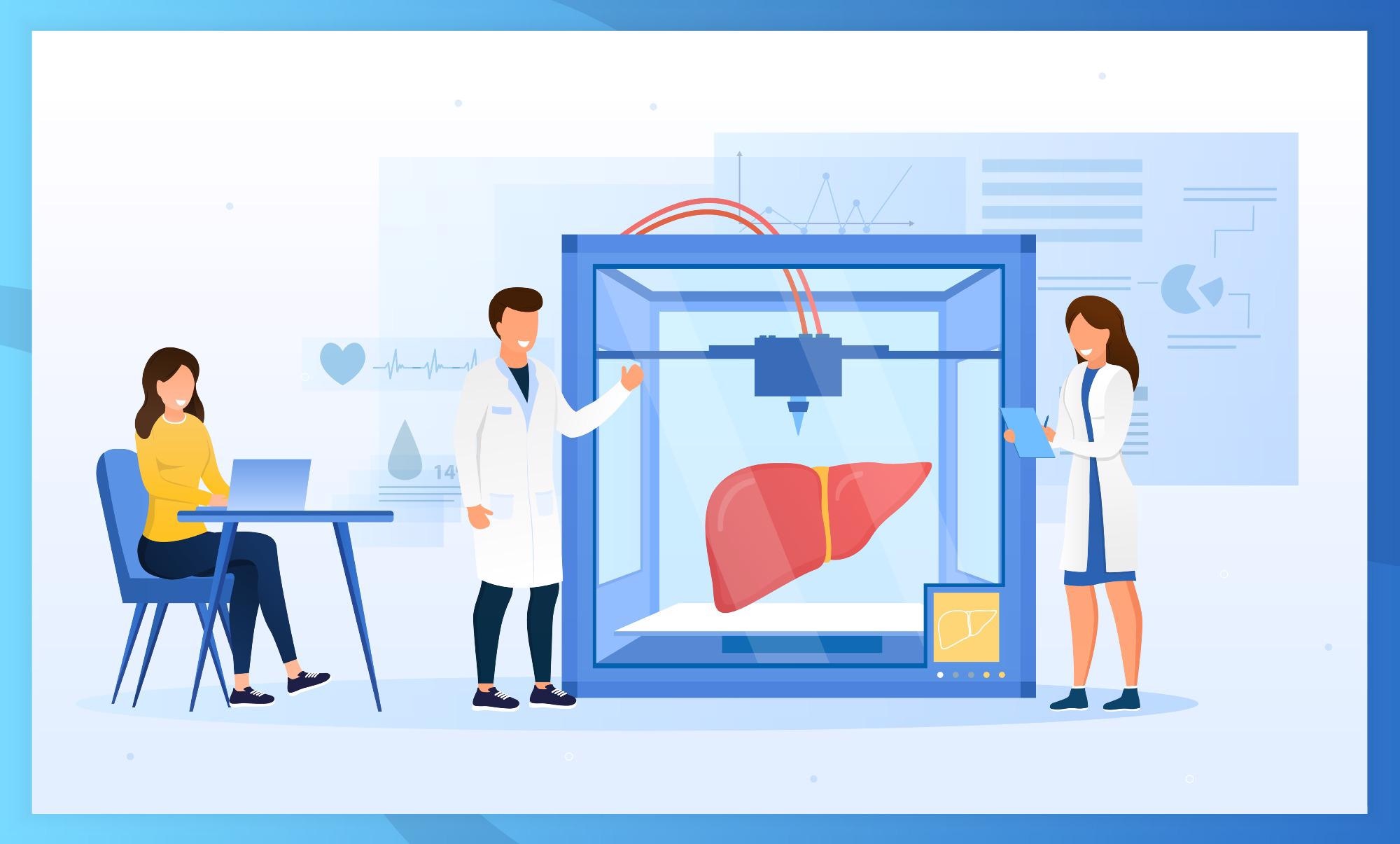
In an article recently published in the journal Bioprinting, researchers discussed the challenges and prospects associated with 3D-bioprinting for mimicking the liver function in micro-patterned units.

Study: Mimicking the liver function in micro-patterned units: Challenges and perspectives in 3D-Bioprinting. Image Credit: Mr.Good/Shutterstock.com
Background
A vast range of diseases can result from any condition affecting the liver's functions. For individuals with liver failure, partial or total organ transplantation has become the gold standard treatment. However, there are several drawbacks to liver transplantation, such as a scarcity of donor organs and poor engraftment success. In this case, liver tissue engineering, which uses the 3D bioprinting process, has provided an alternate approach.
When this technology is combined with suitable cells, printable biomaterials, and growth hormones, a more developed model can be created to better grasp biological intricacies. Bioprinted liver tissue has been proven to overcome present constraints in liver regeneration, liver transplantation, and high throughput drug testing, thanks to recent developments in 3D bioprinting procedures.
With a better understanding of liver microstructure and microenvironment, cellular phenomena, stem cell differentiation technologies, and design and fabrication methodologies, the field of liver bioprinting and tissue engineering has progressed significantly over the last few decades.
About the Study
In this study, the authors discussed the potential of bioprinting three-dimensional (3D) micro-patterned structures for the simulation of complex organs in the body and to generate micro-and macroscale bioengineered structures which resemble the original tissues as much as possible.
Numerous challenges in hepatic tissue engineering such as high vascularization of liver tissue, issues cultivating hepatocytes, and the liver's complicated microenvironment, were discussed. The current obstacles in the bioprinting technology for liver tissue were highlighted along with prospective solutions, and the most appropriate cell types, bioprinters, and hydrogels were also illustrated.
The team discussed the present challenges and limitations of 3D bioprinting in terms of diverse cell sources, varied matrices, bioprinters, and vascularization-related issues in this context. They also determined the main parameters to consider when choosing a hydrogel for bioprinting.
Observations
Several studies showed that various hepatoma cell lines, such as HepG2, Huh7, and HepaRG, could execute a wide range of primary human hepatocytes (PHH) functions. Chaotic flow printing allowed for a high printing speed and a resolution of 10 µm over a 10 cm2 area. Bio-inks with the maximum cell density of up to 1 x 10⁸ – 1 x 10⁹ cells/mL or spheroids could be used with laser-assisted bioprinting, which ensured the production of well-organized liver-like units. A gelatin solution's sol-gel transition temperature was around 28°C, which made it a reliable natural bio-ink for 3D organ bioprinting. Natural polymers were preferred for bioprinting and soft tissue engineering, including the liver, which had a stiffness of 1.5–2 kPa.
Many studies demonstrated that 3D bioprinting could be utilized to improve a biological alternative as a development tool. The application of more consistent materials, cells, and precise bio-bioprinters could be considered important factors in the design and development of a bioprinting technology to better imitate these qualities.
In the case of bioprinters, 4D bioprinting technology was developed as a powerful platform that had given 3D bioprinting a "time value." This method employed stimuli-responsive (smart) biomaterials capable of modifying the geometry of a construct over time, which allowed the creation of more complicated structures. When compared to traditional 3D bioprinting systems, the 4D bioprinting approach resulted in a more satisfying microenvironment design. In terms of cell sources, stem cell-derived hepatic-like cells (HLCs) appeared to be more promising than primary hepatocytes for the development of 3D structures.
Furthermore, due to the difficulty of imitating real liver functional qualities and cell density, 3D printed liver structures containing individual cells had limitations. Bioprinting of spheroids was used in numerous studies to overcome this problem, and bio-inks were the most crucial component for effective bioprinting. Furthermore, sufficient rigidity was essential to keep the entire structure stable over time.
Conclusions
In conclusion, this study elucidated that while 3D bioprinting is still in its early stages, it has the potential to overcome many of the obstacles associated with the production of complex tissues in order to simulate natural conditions in the human body. The authors emphasized that bio-inks made up of novel biomaterials combined with decellularized extracellular matrix (dECM) could be excellent for bioprinting. By substituting a 3D structure for a monolayer culture, the constraints in liver tissue engineering can be addressed.
The team believes that in future research efforts, balancing the bio-printability and bio-functionality of bio-inks should be considered. Also, to create multi-component bio-inks with the same structure as the liver ECM, a thorough understanding of the liver ECM is required. Multicomponent bio-inks have been studied in conjunction with the liver dECM in a number of investigations.
Overall, the findings of this study suggest that a uniform decellularization procedure that can entirely remove cellular components while preserving ECM elements must be proposed and validated. It is also critical to use unique cross-linking techniques to quickly generate biocompatible printable multicomponent bio-inks.
More from AZoM: How are Micrometers Used to Measure Surface Metrology?
Disclaimer: The views expressed here are those of the author expressed in their private capacity and do not necessarily represent the views of AZoM.com Limited T/A AZoNetwork the owner and operator of this website. This disclaimer forms part of the Terms and conditions of use of this website.
Source:
Heydari, Z., Pooyan, P., Bikmulina, P., et al. Mimicking the liver function in micro-patterned units: Challenges and perspectives in 3D-Bioprinting. Bioprinting e00208 (2022). https://www.sciencedirect.com/science/article/pii/S2405886622000185