The Hitachi TM4000PlusIII scanning electron microscope is a compact and user-friendly solution for imaging and elemental analysis. This innovative tabletop SEM, designed for a variety of applications ranging from research labs to industrial quality control, provides dependable imaging capabilities without the price and space constraints of a standard SEM.
Product Features
- Automation ensures consistent and dependable imaging
- Imaging with high resolution, depth-of-focus, and multiple modes
- Elemental analysis is optional
- Navigate with ease utilizing optical camera integration
- Suitable for conductive and non-conductive samples
- Maintenance planning with filament monitoring
Specifications
- Magnifications: 25×–250,000× (monitor display); 10×–100,000× (photography)
- Voltage Acceleration: 5, 10, 15, 20 kV
- Probe Current Mode: 5 steps
Why Choose the TM4000Pluslll?
Whether the user is a research scientist hoping to enhance their understanding of materials or a quality control manager seeking consistent results, the TM4000PlusIII is designed to satisfy their demands.
This microscope combines the capabilities of scanning electron microscopy with a convenient tabletop form. It is easy to use and offers excellent imaging, navigation, and analytic capabilities.
With automated functionality to expedite repetitive processes, a high signal-to-noise ratio for clear imaging, and an easy-to-use interface, this SEM makes electron microscopy more accessible and productive than ever.

Image Credit: Hitachi High-Tech Europe
Features and Benefits
Automation for Consistent and Reliable Imaging
- Auto-focus, brightness, and contrast provide consistent results in multi-user scenarios
- Inexperienced users can operate more easily thanks to automated stage movement, magnification changes, and image capture
- Reproducible workflows without the need for coding knowledge are made possible by optional tools like Multi-Zigzag, Python scripting, and EM-Flow Creator

Image Credit: Hitachi High-Tech Europe
Well-Resolved, High Depth-of-Focus and Multiple-Mode Imaging
- Precise, contrast-rich imaging at 5, 10, or 15 kV
- Surface, topographical, and compositional information is obtained using secondary and backscattered electron detectors
- A single image with mixed output from both detectors provides thorough imaging insights

Image Credit: Hitachi High-Tech Europe
Enhanced Imaging Flexibility and Elemental Analysis
- Optional integrated EDX for material composition analysis using 30 mm or 60 mm sensors
- For detailed sample examinations, switch effortlessly between imaging and elemental mapping
- Compatible with AZtecLiveLite, which allows for efficient and high-throughput analysis

Image Credit: Hitachi High-Tech Europe
Navigate with Ease using Optical Camera Integration
- The integrated high-resolution optical camera creates a zoomable map overview of samples
- Combine optical images with SEM snapshots for accurate navigation and smooth operation

Image Credit: Hitachi High-Tech Europe
Ideal for Educational and Non-Conductive Samples
- Low-vacuum functionality allows for clear imaging of sensitive or non-conductive materials that do not require substantial preparation
- Suitable for educational contexts, students and early-career researchers can readily investigate electron microscopy

Image Credit: Hitachi High-Tech Europe
Maintenance Planning with Filament Monitoring
- Tracks filament usage and calculates remaining lifespan for proactive maintenance planning
- Reduces downtime and guarantees continuous processes in labs with numerous users

Image Credit: Hitachi High-Tech Europe
Applications Gallery
Metals
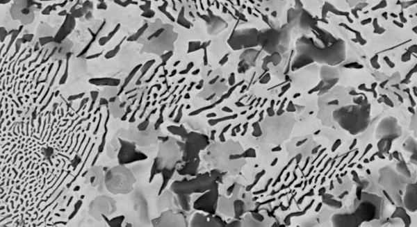
White metal. Acceleration voltage: 20 kV, Magnification: 2,000x. Image Credit: Hitachi High-Tech Europe

Corroded copper wire. Acceleration voltage: 10 kV, SE signal. Image Credit: Hitachi High-Tech Europe
Chemistry

STEM observation of grease thickener. Magnification: 9,000x. Image Credit: Hitachi High-Tech Europe

STEM observation of grease thickener. Magnification: 10,000x. Image Credit: Hitachi High-Tech Europe
Biology

Rat blood vessels (deparaffinized sections). Acceleration voltage: 15 kV, Magnification: 800x. Image Credit: Hitachi High-Tech Europe

Rat blood vessels (deparaffinized sections).Acceleration voltage: 15 kV, Magnification: 3,000x. Image Credit: Hitachi High-Tech Europe
Specifications
Source: Hitachi High-Tech Europe
| . |
. |
| Magnifications |
10x - 100,000x (Photographic)
25x - 250,000x (Monitor display) |
| Accelerating Voltage |
5 kV, 10 kV, 15 kV, 20 kV |
| Probe Current Mode |
5 steps |
| Image Signal |
Backscattered, Secondary, Mixed |
| Vacuum Mode |
Conductor: BSE, CL
Standard: BSE/SE/Mixed
Charge-up reduction:
BSE/SE/Mixed |
| Stage Travel Range |
X: 40 mm, Y: 35 mm |
| Camera Navigation System |
Included |
| Filament Type |
Pre-centered cartridge tungsten hairpin |
| Signal Detection System |
High-Sensitivity 4-segment BSE,
Low-Vacuum SE / CL |
| Auto Image-Adjustment |
Auto start, focus, brightness |
| Image Data Saving |
2560 x 1920 pixels, 1280 x 960 pixels, 640 x 480 pixels |
| Evacuation System |
Turbo molecular pump, diaphragm pump |
| Operation Help Functions |
Raster rotation, magnification presets, image shift |
| Safety Functions |
Over-current protection, built-in ELCB |