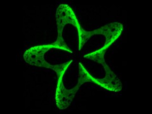
A new technique called 3D photografting in which laser beams are employed to position molecules in three dimensional materials has been developed by scientists at the Vienna University of Technology.
 3D Pattern from Photografting
The technique can be employed to develop sensors at the micro scale or to grow biological tissues. While there are numerous methods to engineer three-dimensional objects at the micrometer level, it has been difficult to control the material’s chemical properties at the micro scale. The new technique of positioning molecules will enable the positioning of chemical signals during tissue growth in the body to indicate the place where living cells can attach.
The scientists used a material called hydrogel, which is composed of macromolecules set in a loose mesh. The pores in between the molecules allow the passage of other molecules and also cells. The scientists introduced specially chosen molecules into the mesh and irradiated specific points using a laser beam. Photochemical labile bonds were split at the spots where the laser beam was very intense. This led to the generation of extremely reactive intermediary molecules that bonded with the hydrogel rapidly. The precision with which the molecules are positioned is dependent on the laser resolution.
For tissue growth in the human body, cells need a scaffold to hold on to. The scaffold or the place the cells are to grow is indicated to the cells through certain amino acid sequences. Researchers replicate these signals to artificially grow tissues on two-dimensional surfaces. However, engineering larger tissues require a three-dimensional technique similar to the 3D photografting. The technique has applications not only in bioengineering but also in sensor and photovoltaic technologies.