Feb 22 2013
When a shark is spotted in the ocean, humans and marine animals alike usually flee. But not the remora – this fish will instead swim right up to a shark and attach itself to the predator using a suction disk located on the top of its head.
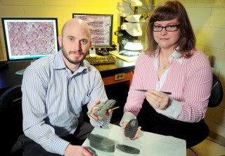
 GTRI senior research engineer Jason Nadler (left) and GTRI research scientist Allison Mercer display 3-D rapid prototypes of the remora's adhesive disk, which the fish uses to attach and detach from sharks and other hosts. Behind the researchers, computer screens show a scanning electron microscope image of the scales on a mako shark (left) and an optical micrograph of a remora's adhesive disk, which revealed similar spacing between features. Georgia Tech Photo: Gary Meek
While we know why remoras attach to larger marine animals – for transportation, protection and food – the question of how they attach and detach from hosts without appearing to harm them remains unanswered.
A new study led by researchers at the Georgia Tech Research Institute (GTRI) provides details of the structure and tissue properties of the remora’s unique adhesion system. The researchers plan to use this information to create an engineered reversible adhesive inspired by the remora that could be used to create pain- and residue-free bandages, attach sensors to objects in aquatic or military reconnaissance environments, replace surgical clamps and help robots climb.
“While other creatures with unique adhesive properties – such as geckos, tree frogs and insects – have been the inspiration for laboratory-fabricated adhesives, the remora has been overlooked until now,” said GTRI senior research engineer Jason Nadler. “The remora’s attachment mechanism is quite different from other suction cup-based systems, fasteners or adhesives that can only attach to smooth surfaces or cannot be detached without damaging the host.”
The study results were presented at the Materials Research Society’s 2012 Fall Meeting and will be published in the meeting’s proceedings. The research was supported by the Georgia Research Alliance and GTRI.
The remora’s suction plate is a greatly evolved dorsal fin on top of the fish’s body. The fin is flattened into a disk-like pad and surrounded by a thick, fleshy lip of connective tissue that creates the seal between the remora and its host. The lip encloses rows of plate-like structures called lamellae, from which perpendicular rows of tooth-like structures called spinules emerge. The intricate skeletal structure enables efficient attachment to surfaces including sharks, sea turtles, whales and even boats.
To better understand how remoras attach to a host, Nadler and GTRI research scientist Allison Mercer teamed up with researchers from the Georgia Tech School of Biology and Woodruff School of Mechanical Engineering to investigate and quantitatively analyze the structure and form of the remora adhesion system, including its hierarchical nature.
Remora typically attach to larger marine animals for three reasons: transportation – a free ride that allows the remora to conserve energy; protection – being attacked when attached to a shark is unlikely; and food – sharks are very sloppy eaters, often leaving plenty of delectable morsels floating around for the remora to gobble up.
But whether this attachment was active or passive had been unclear. Results from the GTRI study suggest that remoras utilize a passive adhesion mechanism, meaning that the fish do not have to exert additional energy to maintain their attachment. The researchers suspect that drag forces created as the host swims actually increase the strength of the adhesion.
Dissection experiments showed that the remora’s attachment or release from a host could be controlled by muscles that raise or lower the lamellae. Dissection also revealed light-colored muscle tissue surrounding the suction disk, indicating low levels of myoglobin. For the remora to maintain active muscle control while attached to a marine host over long distances, the muscle tissue should display high concentrations of myoglobin, which were only seen in the much darker swimming muscles.
“We were very excited to discover that the adhesion is passive,” said Mercer. “We may be able to exploit and improve upon some of the adhesive properties of the fish to produce a synthetic material.”
The researchers also developed a technique that allowed them to collect thousands of measurements from three remora specimens, which yielded new insight into the shape, arrangement and spacing of their features. First, they imaged the remoras in attached and detached states using microtomography, optical microscopy and scanning electron microscopy. From the images, the researchers digitally reconstructed each specimen, measured characteristic features, and quantified structural similarities among specimens with significant size differences.
Detailed microtomography-based surface renderings of the lamellae showed a row of shorter, more regularly spaced and more densely packed spinules and another row of longer, less densely spaced spinules. A quantitative analysis uncovered similarities in suction disk structure with respect to the size and position of the lamellae and spinules despite significant specimen size differences. One of the fish’s disks was more than twice as long as the others, but the researchers observed a length-to-width ratio of each specimen’s adhesion disk that was within 16 percent of the average.
Through additional experiments, the researchers found that the spacing between the spinules on the remoras and the spacing between scales on mako sharks was remarkably similar.
“Complementary spacing between features on the remora and a shark likely contributes to the larger adhesive strength that has been observed when remoras are attached to shark skin compared to smoother surfaces,” said Mercer.
The researchers are planning to conduct further tests to better understand the roles of the various suction disk structural elements and their interactions to create a successful attachment and detachment system in the laboratory.
“We are not trying to replicate the exact remora adhesion structure that occurs in nature,” explained Nadler. “We would like to identify, characterize and harness its critical features to design and test attachment systems that enable those unique adhesive functions. Ultimately, we want to optimize a bio-inspired adhesive for a wide variety of applications that have capabilities and performance advantages over adhesives or fasteners available today.”