Mar 19 2015
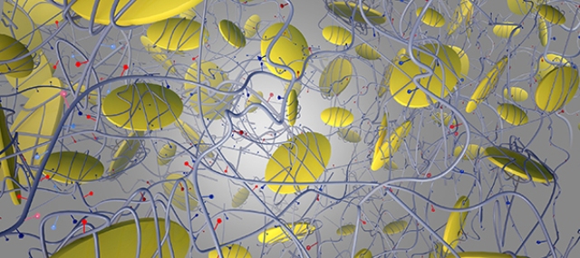
 Tiny nanosilicate particles that are 100,000 times thinner than a sheet of paper are embedded in a collagen-based hydrogel, forming a material that helps trigger bone formation within the body.
Researchers from the Texas A&M University have created a new material that stimulates the stem cells to generate bone cells thereby effectively treating hard-to-heal bone damages and defects.
Akhilesh Gaharwar, assistant professor of biomedical engineering at Texas A&M, said that the research could help change the approach of medical personnel towards treating bone fracture that takes time to heal, and also other bone graft procedures.
The study is published in the “ACS Nano” journal and funded by the National Institutes of Health and the National Science Foundation.
The new biomaterial consists of a gel containing nano-sized, two-dimensional particles which trigger bone growth via a complex signaling mechanism. The mechanism does not involve the use of growth factors that are employed in traditional treatment. Growth factors, when used in large quantities to stimulate cells, can result to serious side effects.
“We are trying to overcome these problems by avoiding the use of growth factors as we recapitulate the natural bone-healing process. Our material is a totally different, alternative strategy in which by using minerals we can induce differentiation in stem cells and promote formation of bone-like tissue, ” Gaharwar said.
Gaharwar explained that these minerals contain large quantities of orthosilicic acid, magnesium and lithium combined with small nanosilicate particles 100,000 times thinner than a paper sheet. These ultrathin nanoparticles are coated with a collagen-based hydrogel that is used in a number of biomedical applications.
The physical, chemical and biological properties of the hydrogel are improved while incorporating the nanosilicates into a gelatin matrix. For instance, the hydrogel can be designed by controlling the interaction between the gelatin and nanosilicates, in order to maintain it at the site of injury for specific durations. This enables the injected hydrogel to enter the defect cavity to heal the site followed by slow degradation in the tissue.
Analysis of the material's mechanical properties is also challenging. Upon injecting into the body, the stiffness of the material is increased by three-to-four times which allows the material to be locked in a place. This stops the material from flowing to the other parts, preventing any side effects.
Gaharwar said that the obtained results were positive, which was indicated by the short-term and long-term indicators of bone growth. Initial tests indicate an increase in alkaline phosphatase activity by three times, which means the signaling process has started to trigger the stem cells for bone cell differentiation. However, the positive results of the late markers indicate increase in the presence of calcium phosphate in bone, by four times.
“The dynamic and bioactive nanocomposite gels we have developed show strong promise in bone tissue engineering applications,” Gaharwar said.
Further research involves an in-depth investigation of the process of nanoplatelets trigger cell differentiation. The shear-thinning property of gel can be used to print three-dimensional tissue structures loaded with cells. Based on this characteristic, Gaharwar and his colleagues are on the verge of developing customized, vascularized scaffolds with the material for insertion into the injury site.
The scaffolds allow the supply of blood to the injury site, resulting to the enhanced healing process initiated by the nanoparticles. The researcher believes that this solution will address the key challenges related to analysis of complex tissues or organs.
“Based on our strong preliminary studies, we predict that these highly biofunctional particles have immense potential to be used in biomedical applications,” Gaharwar added.
The co-workers of the research include Elizabeth Cosgriff-Hernandez and Roland Kaunas, associate professors in the Department of Biomedical Engineering at Texas A&M