Jan 21 2021
In a breakthrough achievement that needs cutting-edge technology, perseverance, and a great deal of caution, researchers have effectively utilized powerful X-rays to investigate irradiated nuclear fuel.
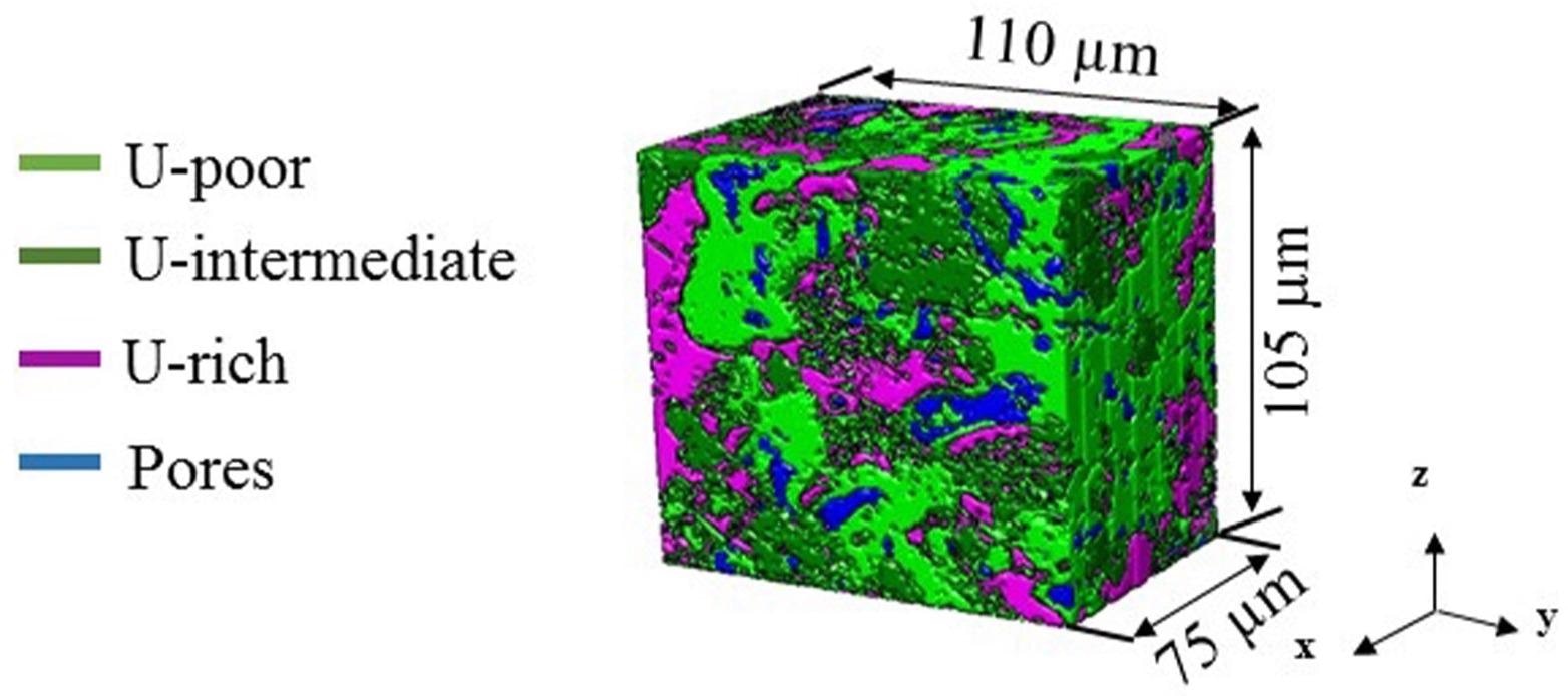
 3D image reconstruction of a sample of irradiated fuel, showing the three thresholded uranium phases co-existing with pores. Image Credit: Maria Okuniewski/Purdue University.
The imaging, headed by investigators from Purdue University and performed at the Argonne National Laboratory of the U.S. Department of Energy (DOE), has disclosed a three-dimensional (3D) view of the inner structure of fuel, laying the basis for more improved designs and models for nuclear fuels.
To date, analyses of uranium fuel have been largely restricted to surface microscopy or to numerous characterization methods using simulated versions that contain negligible radioactivity. However, investigators want to gain a deeper understanding of how the material modifies as it undergoes fission within a nuclear reactor.
The insights, thus gained from this research work, can result in nuclear fuels that not only work more efficiently but also cost less to design. The study was published in the Journal of Nuclear Materials in August 2020.
To visualize the interior structure of the analyzed uranium-zirconium fuel, the team sectioned off a small part of the used fuel that was sufficiently small to be managed safely—a capability that was achieved only within the past seven years.
Then, to get an interior view of this small metallic element, the researchers sought the help of the Advanced Photon Source (APS)—a DOE Office of Science User Facility based at Argonne National Laboratory.
A Study Decades in the Making
But before doing the daunting task of separating a fuel sample and positioning it under an X-ray beam, the researchers needed to identify a suitable sample.
After investigating fuels archived at the Idaho National Laboratory (INL) of the DOE, the team eventually found a uranium-zirconium fuel. This fuel had spent a total of two years and at full power in the Fast Flux Test Facility based in Hanford, Washington, and was taken off the reactor in the early 1990s.
We had to wait decades for this fuel to radiologically cool or decay. It was literally the coolest specimen that we could remove based on the permissible safety guidelines at both INL and APS.
Maria Okuniewski, Study Lead Author and Assistant Professor of Materials Engineering, Purdue University
From the radiological standpoint, even the coolest available specimen of spent fuel was still extremely hot, at its original size. Collected from a larger fuel pin, the specimen measured less than a quarter of an inch in height, but measured 1,200 millirem every hour from a distance of 30 cm—a radiation level that is 240 times more than the permissible limit at the APS.
To minimize the radioactivity level, the investigators produced a relatively smaller specimen using a focused ion beam with scanning electron microscopy at INL. This tool enabled them to pinpoint a target area and deploy a beam of ions that fundamentally milled out a cube of material. The sample, thus obtained, measured around 100 µm across, and was no bigger than the width of a single strand of human hair.
We’ve come a long way with this new instrumentation that allows us to obtain samples that are small enough to be safe and easily handled.
Maria Okuniewski, Study Lead Author and Assistant Professor of Materials Engineering, Purdue University
The tiny sample was placed on a pin, covered in a double-walled tube, and then delivered to the Argonne National Laboratory, with numerous radiation checks to guarantee safety along the way.
At the Argonne National Laboratory, the researchers from Purdue University worked with investigators from beamline 1-ID-E—a high-brilliance X-ray source at the APS—to study the specimen. The objective of the team is to get an interior view of the uranium-zirconium fuel after it has been struck with neutrons for a couple of years.
We are really talking about a piece of dust that you can barely see with the naked eye — it’s that small. But this is also very dense material, so you need a sufficient intensity of high-energy X-rays to penetrate and study it.
Peter Kenesei, Study Co-Author and Physicist, X-ray Science Division, Argonne National Laboratory
The researchers used a method—called micro-computed tomography—that detects the X-ray beam at high resolution as it appears on the other side of the specimen. By using numerous images captured during the rotation of the fuel, computers could rebuild its interior features based on the way it modified the incoming beam, just like a medical CT scan.
“The 1-ID-E beamline’s flexibility, along with Argonne’s expertise in safely handling nuclear materials, allows us to design and conduct a unique experiment like this one,” added Kenesei.
Closer Look at Fuel Swelling
Okuniewski and her collaborators were specifically interested in the phenomenon of swelling. To produce energy, nuclear fuel takes a single uranium atom and splits it into two. This fission process creates byproducts, like the gas xenon, and metals, such as neodymium and palladium. When atoms split and fission products build up, the fuel increases in volume.
The longevity and safety of any specified nuclear fuel relies on the ability to estimate the degree of its swelling. Excess swelling can cause the uranium to react with, and perhaps break down, its outer protective layer, known as cladding. Hence, to stop that from occurring, engineers depend on fuel performance codes, which are essentially computer models that replicate numerous features of a fuel’s behavior in a reactor, like how its components redistribute in space and how hot the fuel will become in temperatures.
“In every single fuel type, swelling is an issue. These fuels are designed so that the inner core is free to expand to a specific level before it touches the cladding,” added Okuniewski.
Apart from offering a clearer and localized picture of the structure of the fuel and the different phases of the material that developed over time, the research work conducted at the APS clearly demonstrated that the emission of fission gases may continue to take place beyond the thresholds believed in earlier studies.
A data of this kind can help reinforce the fuel performance codes, which consequently would further reduce the cost involved in the development of fuels, since consistent computer simulations can reduce the number of costly irradiation tests required.
“We’re always striving within the nuclear community to figure out ways that we can improve the fuel performance codes. This is one way to do that. Now we have three-dimensional insight that we previously didn’t have at all,” Okuniewski concluded.
The study titled, “The application of synchrotron micro-computed tomography to characterize the three-dimensional microstructure in irradiated nuclear fuel,” was funded by DOE’s Office of Nuclear Energy and Purdue University.
Apart from Okuniewski and Kenesei, other authors of the study are Jonova Thomas, Alejandro Figueroa Bengoa, Sri Tapaswi Nori, and Ran Ren from Purdue University; Jon Almer from the Argonne National Laboratory; James Hunter from the Los Alamos National Laboratory; and Jason Harp from the Idaho National Laboratory.
The team would specifically like to acknowledge the efforts of John Vacca, a senior health physicist from the Argonne National Laboratory, who worked to ensure the safety of people during this experiment and several other ones. Vacca has passed away unexpectedly in May 2020.
Scientists gain an unprecedented view of irradiated nuclear fuel
Five pore stages observed in neutron irradiated uranium zirconium fuel. Video Credit: Purdue University College of Engineering.
Journal Reference:
Thomas, J., et al. (2020) The application of synchrotron micro-computed tomography to characterize the three-dimensional microstructure in irradiated nuclear fuel. Journal of Nuclear Materials. doi.org/10.1016/j.jnucmat.2020.152161.