May 21 2021
In the past few years, 3D printing, which is also known as additive manufacturing, has gained momentum.

Image Credit: shutterstock.com/Giovanni Cancemi
The process involves constructing consecutive layers of raw materials like ceramics, plastics and metals, thus offering the main benefit of being able to generate highly complicated geometries or shapes that would be almost impracticable to build via more conventional techniques like molding, grinding, or carving.
The technology provides huge ability in the health care sector. For instance, doctors can employ it to make products to correspond to the anatomy of a patient.
A radiologist can make a precise replica of a patient’s spine to help schedule a surgery; a dentist could scan a broken tooth of a patient to make a completely fitting crown reproduction. The question is what if an additional step has been taken and 3D printing techniques are applied to neuroscience.
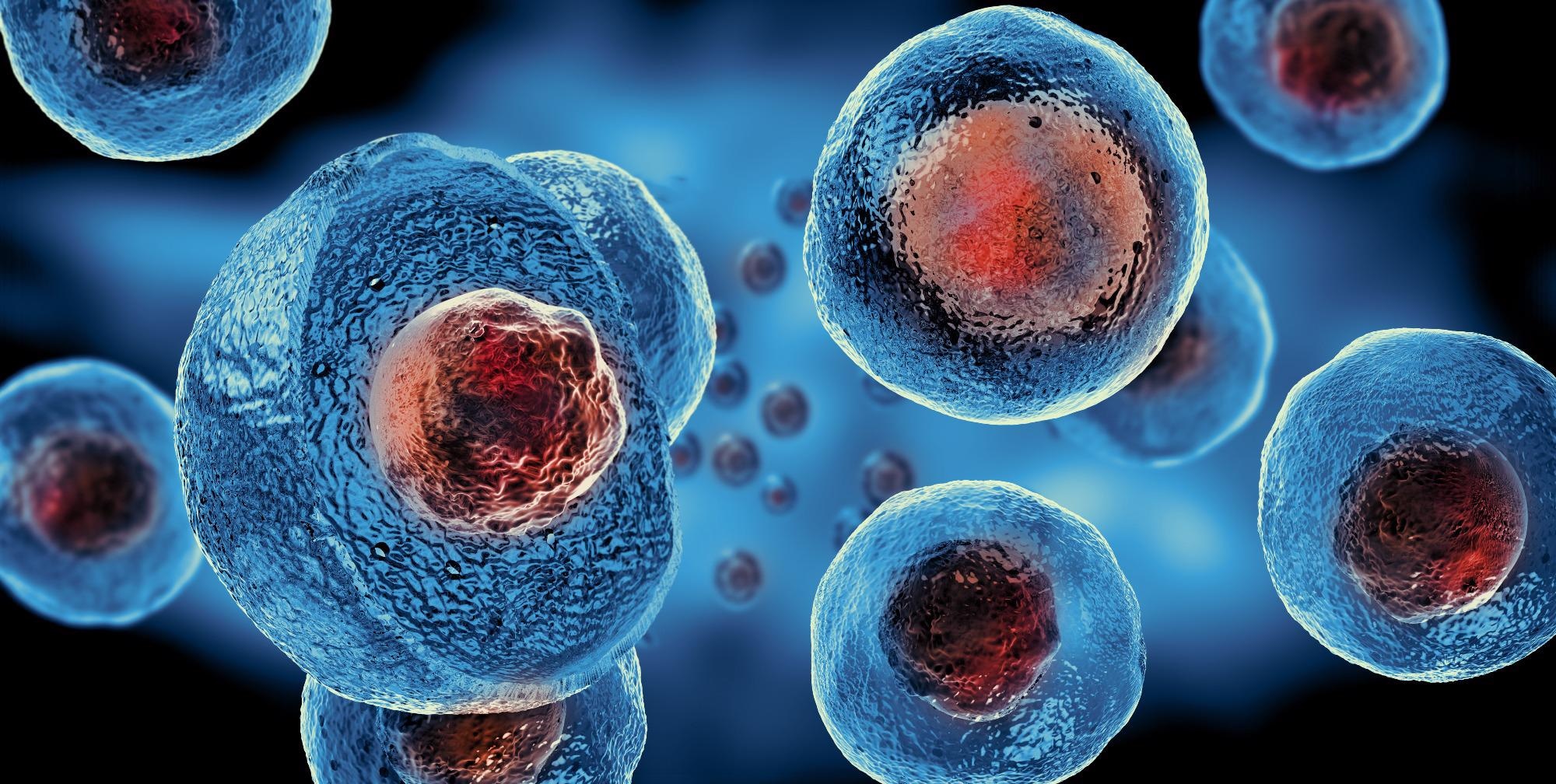
Stem cells are considered as the raw materials of the body. They are pluripotent elements from which all other cells with specialized functions are produced.
The advent of techniques to separate and produce human stem cells has excited many with the guarantee of enhanced human cell function understanding, finally utilizing them for regeneration in trauma and disease.
But the conventional two-dimensional growth of derived neurons—utilizing flat Petri dishes—arises as a significant astonishing factor as it does not sufficiently simulate the myriad developmental cues present in real living organisms, nor in vivo three-dimensional interactions.
To tackle this drawback of present neuronal culturing methods, the FET provided financial support to the MESO-BRAIN project, headed by Aston University.
It suggested an extremely ambitious interdisciplinary enterprise to build truly 3D networks that not only exhibited in vivo activity patterns of neural cultures but also enabled accurate interaction with such cultures.
This enables the activity of separate elements to be readily tracked and regulated via electrical stimulation.
The potential to develop human-induced pluripotent stem cell-derived neural networks upon a specified and reproducible 3D scaffold that can imitate brain activity enables an extensive and elaborate investigation of neural network development.
The MESO-BRAIN project makes it easier to gain better insights into neuronal growth and human disease progression while allowing the advent of large-scale human cell-based assays to check the modulatory effects of toxicological and pharmacological compounds on neural network activity.
Eventually, this can help to better comprehend and treat neurological conditions like trauma, dementia and Parkinson’s disease. Besides, the use of more physiologically appropriate human models will enhance drug screening efficiency and decrease the requirement for animal testing.