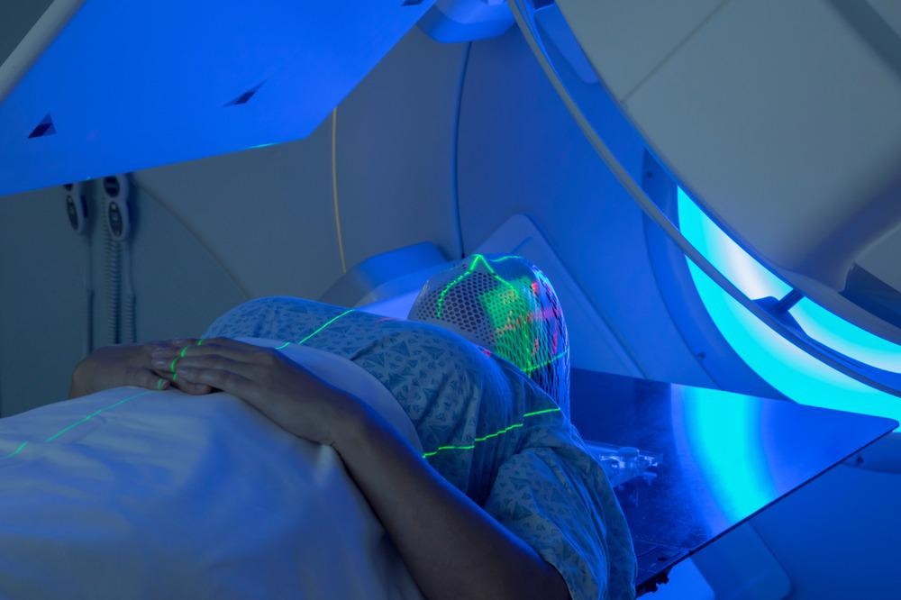
A team of researchers from France has investigated the effects of radiotherapy on tooth enamel and the potential induction of tooth decay. Their findings have appeared in the journal Dental Materials.

Study: The effect of therapeutic radiation on dental enamel and dentin: A systematic review. Image Credit: Mark_Kostich/Shutterstock.com
Background to the Research
Worldwide, head and neck cancers were estimated to have caused 500,000 deaths in 2020. These cancers, including nose, throat, sinus, and mouth cancer are typically treated with radiotherapy. However, it has been observed in numerous studies and medical interventions that following radiotherapy treatments, the onset of tooth decay commonly occurs.
Tooth decay onset typically occurs after a period of four to six months post-treatment. Tooth decay can be caused by hyposalivation and modification of oral flora towards more carcinogenic species of bacteria, or changes in dentin and tooth enamel. Tooth decay can spread rapidly, requiring constant post-treatment observation of patients and medical intervention.
Radiation has an effect on both organic and inorganic matrices. However, due to the low organic content of enamel, quantification of results can be challenging, limiting any conclusions which could be drawn on the effect of irradiation on this tissue type. The collagen matrix is highly susceptible to radiation. Its hierarchical structure is subject to multiple modifications. Several effects have been observed on the inorganic matrices of hard dental tissue.
The effect of radiation on mineralized tooth structures is poorly understood currently. Systematic reviews have been published on the effect of therapeutic radiation on dental tissues in the past decade, highlighting impacts such as extensive prismatic structure damage to teeth and reduced biomechanical properties. Obliteration of tubules has been observed in studies. Different levels of tooth damage have been identified depending on the intensity of delivered radiation doses.
The development of new analytical tools in the past decade has provided new insights into the mechanism of radiation-induced tooth decay, leading to an enhanced understanding of the impact of radiation on the mechanical, structural, and chemical properties of human dentin.
The Study
The study has systematically reviewed the effect of therapeutic radiation used to treat head and neck cancers on tooth decay in patients along with investigation methods over the past decade. The effect of radiation on coronal dentin, dental enamel, and root dentin has been investigated thoroughly by the authors.
A literature search for in vivo and in vitro studies analyzing the effect on human teeth before and after radiation treatment. The authors identified 353 articles with twenty-eight inclusion criteria. Studies that investigated the dentin-enamel junction were excluded as this area of research has been adequately covered in a pre-existing systematic review in 2020. Likewise, studies that exclusively investigated bonding were excluded as this research has been covered by a systematic review in 2017.
Papers were thoroughly investigated for factors such as bias that would limit their veracity. Generally speaking, many of the papers were well-conducted, but some possessed methodological weaknesses and poor understanding of characterization methods, limiting the relevance of their findings. Moreover, few details were given in several studies on their normalization procedures.
Study Findings
The new paper has found current research varies in its means of investigations and parameters evaluated to elucidate the effect of therapeutic radiation on the evolution of mineralized tooth material properties. However, current studies are limited by a lack of consensus.
Studies have demonstrated that irradiation has a significant impact on coronal and root dentin’s organic matrix. Reduced mineral/matrix ratio has an effect on the mineral phase’s crystalline structure. Some studies have indicated that enzymatic action may play a role in organic matrix degradation.
Research has highlighted that decreased crystallinity indexes and hardness values occur in dental enamel exposed to radiation. However, these studies have not correlated this with chemical composition.
There has been a growing interest in characterizing the effects of therapeutic radiation employed to treat head and neck cancer on hard dental tissue. This can be attributed to the development of more powerful analytical techniques in recent years which have allowed researchers to study these effects more thoroughly. However, there is still a lack of a common methodology that hinders effective data comparison.
The authors have stated that research will require a cross-functional and collaborative approach between biologists, dental clinicians, materials scientists, and physicists. This approach will improve the understanding of the effect of radiation on dental tissue and the causes of post-treatment tooth decay. The new paper has provided a solid knowledge base for future studies into how the evolution of post-cancer treatment tooth decay is influenced by the use of therapeutic radiation.
More from AZoM: What are Profile Roughness Parameters?
Further Reading
Douchy, L et al. (2022) The effect of therapeutic radiation on dental enamel and dentin: A systematic review Dental Materials [online, corrected proof] sciencedirect.com. Available at: https://doi.org/10.1016/j.dental.2022.04.014
Disclaimer: The views expressed here are those of the author expressed in their private capacity and do not necessarily represent the views of AZoM.com Limited T/A AZoNetwork the owner and operator of this website. This disclaimer forms part of the Terms and conditions of use of this website.