Mar 15 2016
Using the idea of colorful "grow capsules" in water that blossom into animal-shaped sponges, scientists have created biodegradable polymer grafts. These grafts, when surgically implanted in damaged vertebrae, can grow to the required shape and size in order to fix the spinal column.
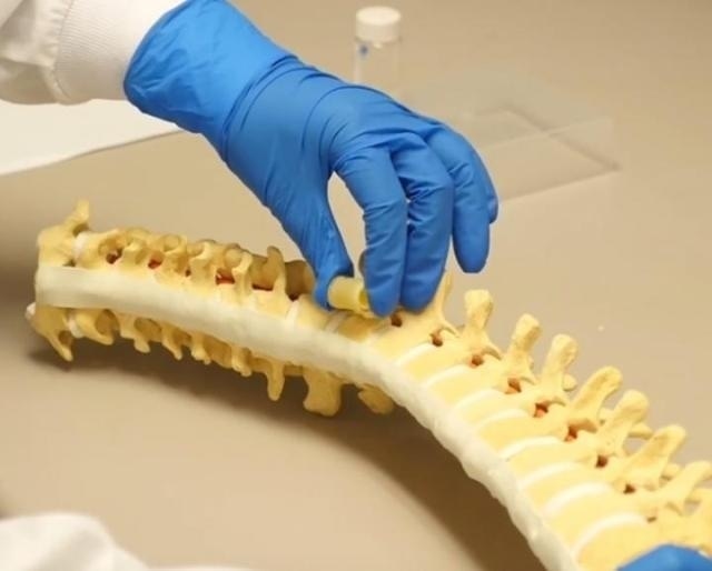
 A polymer bone graft (held above in the top gloved hand) can expand in the body to just the right size to replace excised spinal tissue. (Photo credit: Lichin Lu Video credit: American Chemical Society)
This research work was presented at the American Chemical Society’s (ACS) 251st National Meeting & Exposition. ACS is the world's largest scientific society. The meeting is being held in San Diego till March 17, and features over 12,500 presentations on several science topics. A new video footage on the research work is available online at http://bit.ly/ACSBone.
The overall goal of this research is to find ways to treat people with metastatic spinal tumors. The spine is the most common site of skeletal metastases in cancer patients, but unlike current treatments, our approach is less invasive and is inexpensive.
Lichun Lu, Ph.D.
Usually in the case of extensive spinal tumors, the complete bone segment - as well as nearby intervertebral discs - need to be removed from the damaged area, creating a large void or gap. This gap must be filled to protect the spinal cord and to maintain the spine’s integrity.
Two surgical choices exist in cases of major spinal metastases. The invasive and more aggressive option is where the chest cavity of the patient is opened, to allow sufficient room to insert bone grafts or metal cages as a substitute for the missing fragment. The second less invasive option only needs a small cut in the patient’s posterior, but offers enough space to insert the costly, short expandable titanium rods.
To develop a less expensive graft for the posterior spinal surgery, Lu, who is at the Mayo Clinic, and her postdoctoral fellow, Xifeng Liu, Ph.D., looked for a material that could be dehydrated to a smaller size in tune with the posterior surgery, and after implantation, could absorb body fluids and expand to replace the missing vertebrae.
The researchers began by cross-linking oligo[poly(ethylene glycol) fumarate] to form a hollow hydrophilic cage – the scaffold of the graft – which could later be filled with therapeutics and stabilizing materials.
When we designed this expandable tube, we wanted to be able to control the size of the graft so it would fit into the exact space left behind after removing the tumor.
Lichun Lu, Ph.D.
The scientists also had to control the expansion kinetics. A surgeon may not find sufficient time to position the cage correctly if it enlarges rapidly, but a slow enlargement would result in a longer surgery time.
Manipulating the extent and the time needed for the expansion of the polymer graft involved knowledge of the polymer’s chemistry,
By modulating the molecular weight and charge of the polymer, we are able to tune the material's properties.
Lichun Lu, Ph.D.
The scientists analyzed the effects caused by the chemical changes by monitoring the grafts' rate of expansion under simulated spinal column environments in the lab. This information is important in order to find out the optimal size of the graft implant for the restorative spinal surgery. The team decided on a combination of materials that have been found to be biocompatible in animals, and that they think will also work in humans.
According to Lu, her lab's next step is analyzing the polymer grafts in cadavers, and simulating an in-patient surgery. Their objective is to start clinical trials within a few years.
Lu’s research received funds from the National Institutes of Health.