May 6 2016
Proteins are complex molecules that are responsible for carrying out all of the processes that sustain life. A better understanding of how these molecules perform their functions can be achieved by learning the assembly of their atoms and how the structure of this assembly changes as the atoms react. Until now, no imaging technique has allowed the observation of molecular movement in such speed and detail.
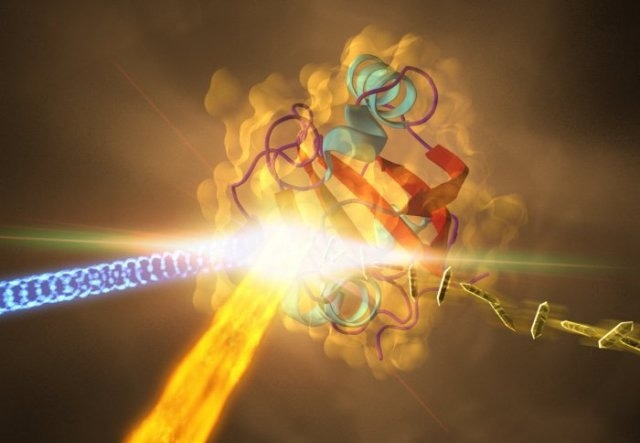
 Interaction between the molecule and light measured with an X-ray beam. (Credit: SLAC National Accelerator Laboratory)
For the first time a group of physicists and biochemists, headed by Imperial College London and the University of Wisconsin-Milwaukee, have recorded the basic processes of chemical reactions occurring in real time. The researchers captured images of a small crystallized protein as it responded to light, all at a speed of 25 trillion per second. Using this approach, the team created an exact picture of the protein activity occurring every few femtoseconds - quadrillionths of a second. The results of the study have been reported in the Science journal.
Traditionally, researchers have relied on a technique known as X-ray crystallography, which is only capable of capturing the static images of proteins. It is now possible to create a set of crystallographic images and convert them into a molecular movie over very short timescales.
Usually, we can only image the structure after the reaction, and infer what has happened. This is the first time we have been able to image crystal structures on timescales where the proteins are still undergoing the reaction. What happens during these timescales determines the outcome of the reaction, so knowing exactly what’s going on is vital. Previously our information and images of how the reactions work have been based on theory and spectroscopy. Now we can see it in reality.
Dr Jasper van Thor, Department of Life Sciences, Imperial College London
Responding to Light
The researchers went on to investigate a yellow dye molecule present at the core of a light-sensitive protein. This protein experiences a shape shift when interacting with a photon, which is nothing but a particle of light. They observed that the fundamental biological process is analogous to how the retina of the human eye reacts to light. With the aid of the Linac Coherent Light Source X-ray Free Electron Laser in California, the researchers fired highly bright pulses at the protein and at the same time captured images every few femtoseconds as the photon reaction advanced. Most importantly, the result was obtained through meticulous customization of the visible light pulse, which is essential for highly bright and short pulses.
We are working in interesting regimes that are new to crystallography, where the properties of the visible pulse matter most.
Dr Jasper van Thor, Department of Life Sciences, Imperial College London
To actively regulate the dynamics, the team will explore ways to obtain femtosecond detail and these could eventually enable the research community to intervene in the functional process of proteins by utilizing light.
Chemistry for All Life
The method has been shown to be effective, so the researchers are hoping that it will soon be used across molecular biology to reveal the mechanisms of proteins’ key reactions for life.
This puts us dramatically closer to understanding the chemistry necessary for all life. Discovering the step-by-step process of how proteins function is necessary not only to inform treatment of disease, but also to shed light on the grand questions of biology.
Marius Schmidt, Physics Professor, UW-Milwaukee
Dr van Thor is exploring other areas and applications where this technology could potentially lead to novel breakthroughs. He is also assisting in the development of a case for a parallel instrument center to be constructed in the UK.
The study was conducted in partnership with institutions from Europe and the US, including the University of Wisconsin-Milwaukee; University of Hamburg; Lawrence Livermore National Laboratory; State University of New York, Buffalo; the Max Planck Institute for Structure and Dynamics of Matter, and University of Jyvaskyla.