Feb 19 2018
Scientists at the Queen Mary University of London have devised an innovative printing method by using molecules and cells usually seen in natural tissues to develop constructs that look similar to biological structures.
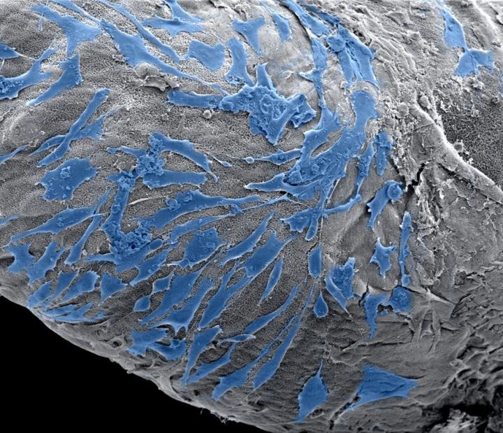
 These are cells spreading on the outside of a PA-based scaffold. (Image credit: Clara Hedegaard)
These structures are integrated into an ink which resembles their native environment and renders it probable to make them function the same way they would function inside the human body.
This enables the scientists to investigate the way cells function within such environments and prospectively allows them to test biological scenarios such as those in which cancer cells grow or the way immune cells interact with normal cells, which can result in the development of innovative drugs.
The method integrates molecular self-assembly, constructing structures by arranging molecules similar to Lego pieces, by adopting additive manufacturing, like 3D printing, to re-develop the complicated structures.
The structures can be produced through digital control and with molecular accuracy, which also allows the scientists to develop constructs that resemble tissues or body parts for regenerative medicine or tissue engineering.
The research has been reported in the journal Advanced Functional Materials.
According to Professor Alvaro Mata from Queen Mary’s School of Engineering and Materials Science, “The technique opens the possibility to design and create biological scenarios like complex and specific cell environments, which can be used in different fields such as tissue engineering by creating constructs that resemble tissues or in vitro models that can be used to test drugs in a more efficient manner.”
The method combines the micro- and macroscopic regulation of structural characteristics that printing offers, with the molecular and nano-scale regulation enabled by self-assembly. Due to this, it attends to a major requirement in 3D printing in which usually used printing inks have restricted potential to vigorously activate the cells to be printed.
This method enables the possibility to build 3D structures by printing multiple types of biomolecules capable of assembling into well-defined structures at multiple scales. Because of this, the self-assembling ink provides an opportunity to control the chemical and physical properties during and after printing, which can be tuned to stimulate cell behavior.
Clara Hedegaard, Ph.D. Student and Lead Author
The research was performed in partnership with the Nanyang Technological University in Singapore and the University of Oxford.
The European Research Council’s Starting Grant (STROFUNSCAFF), the FP7-PEOPLE-2013-CIG Biomorph, the Royal Society, and the European Space Agency’s Drop My Thesis program 2016 supported the study.