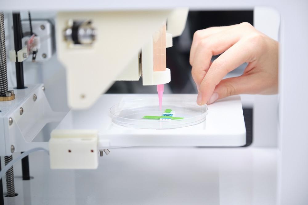
In an article published in the open-access journal Bioprinting, researchers developed a leaf-like channeled device for cell delivery using 3D-bioprinting and computer-aided design (CAD) software and simulated the fluid flow through the channels.

Study: Design and biofabrication of a leaf-inspired vascularized cell-delivery device. Image Credit: Ladanifer/Shutterstock.com
The leaf-like device was 3D-printed using hydrogel made from a mixture of alginate and tunicate cellulose nanofibers crosslinked in calcium chloride (CaCl2) solution, and the flow channels were made up of Pluronic F-127 ink. Additionally, endothelial tissues were formed (i.e. endothelialized) on its surface to overcome size limitations owing to oxygen diffusion. Endothelial cell adhesion in the channels was further enhanced by surface modification through periodate oxidation and laminin conjugation.
Background
Many critical medical conditions can be resolved by artificial organs, which require the maximum utilization of tissue engineering. One of the major hurdles in fabricating larger 3D tissue structures is the proper design and distribution of the vascular system. It consists of a large number of macro-and microchannels for mass transportation of oxygen, nutrients, hormones, and many more. In general, the distance between any cell and the nearest vascular channel should be less than 200 µm.
Devices containing endothelialized vascular networks can be used for stem cell delivery and artificial pancreas for insulin delivery, in case of diabetes. These cell delivery devices consist of an embedded scaffold on a vascular channel network. Additionally, the templates of vascular channels can be 3D-printed using various bio-inks. 3D printing supports the fabrication of thin delicate structures with on-demand customization.
Cellulose is the most abundant biopolymer in the world, which is the main constituent of the cell walls of plants and animals. It can be used as a scaffold material for cell delivery devices. A mixture of crosslinked cellulose nanofibers and alginate can be used as a bio-ink for 3D-bioprinting.
However, cellulose has very poor adhesion to endothelial cells. Thus, it requires chemically induced surface modification to form conjugates with the protein present in the cells and form a durable endothelialized scaffold.
About the Study
In this study, researchers fabricated a leaf-like cell delivery system consisting of a hydrogel-based vascular channel network, a scaffold to host the vascular network, and endothelialization on the scaffold for the diffusion of oxygen and nutrients.
First, the vascular channel network was designed by CAD software and fabricated by Pluronic F-127 ink in a 3D-bioprinting. Then, a leaf-shaped mold of white resin was made using the 3D printer. The homogeneous hydrogel mixture of 20:80 vol% transparent alginate and tunicate nanocellulose was prepared and mixed with lipoaspirate (i.e. adipose tissue) obtained from the abdominal subcutaneous tissue of a human.
More from AZoM: What Raw Materials Can Be 3D Printed?
Subsequently, the tissue-hydrogel mixture was poured onto both sides of the leaf-shaped mold one after another and flipped several times to obtain the adipose-hydrogel leaves. Further, CaCl2 solution was added to it and incubated overnight at 4 ⁰C to facilitate crosslinking and form the final leaf-like device. Finally, human umbilical vein endothelial cells were cultured on the surface of the device in an endothelial growth medium (EGM).
Observations
The color liquid capillary test confirmed the interconnectivity of all side channels to the main channel. Cell vitality was studied after the first and fourth day, which showed that the adipose-derived stem cells (ASC), mixed with the nanocellulose-alginate hydrogel, effectively survived after the casting process of the leaf. However, the cells inside the channels died in large numbers due to the restricted flow of nutrients.
Moreover, during the process of cellulose surface modification for improved adhesion to endothelial cells, sodium periodate oxidation of cellulose created dialdehyde cellulose. The aldehyde groups of dialdehyde cellulose are anchored to the cells by covalently bonding with the proteins of the cells. Due to this conjugation of the hydrogel with the proteins, a robust and durable endothelialization was achieved.
The flow simulation analyzed the effect of the sheer force of the water on cell attachment to the walls of the vascular network. It showed that the flow velocity was the highest for the central channel, and some of the lateral channels suffered from some unwanted recirculating regions and creeping flow. Thus, there was a requirement for further geometrical optimization of the flow channels.
Conclusions
To conclude, the researchers of this study designed and fabricated an artificial cell to deliver a device with a leaf-like vascular channel flow network using CAD software and 3D-bioprinter. The vascular network was made from Pluronic F-127 sacrificial ink and covered by a scaffold made up of adipose tissues containing a nanocellulose-alginate hydrogel mixture.
Further, endothelial tissues were grown on the device surface after surface modification of the cellulose with improved adhesion to simulate a real human organ. The fabrication process demonstrated excellent survivability of the adipose tissues with an efficient, interconnected flow network. However, the study suggests further optimization of flow channels to reduce the shear force of flowing liquids.
Disclaimer: The views expressed here are those of the author expressed in their private capacity and do not necessarily represent the views of AZoM.com Limited T/A AZoNetwork the owner and operator of this website. This disclaimer forms part of the Terms and conditions of use of this website.
Source:
Sämfors, S., Niemi, E., Oskarsdotter, K., Egea, C., Mark, A., Scholz, H., Gatenholm, P., Design and biofabrication of a leaf-inspired vascularized cell-delivery device, Bioprinting, 2022, e00199, ISSN 2405-8866, https://www.sciencedirect.com/science/article/pii/S2405886622000094