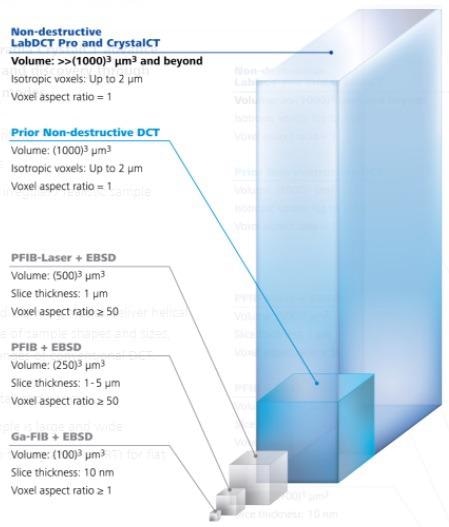
If users wish to execute non-destructive mapping of grain morphology in 3D to describe materials like metals, ceramics or alloys, they can find out the first commercially available laboratory-based diffraction contrast tomography (DCT) method for entire three-dimensional imaging of grains in the sample.
Two strong solutions — LabDCT Pro and CrystalCT — enable users to directly envision the 3D orientation of the crystallographic grain. Programmed with the advanced GrainMapper3D software, it sets the stage to examine a range of polycrystalline materials.
Applications
ZEISS Xradia Versa LabDCT Pro and Xradia CrystalCT develop materials modeling, characterization and breakthrough via ground-breaking diffraction scanning modes.
- Allows scanning bigger sample volumes
- Offers unparalleled sample representivity
- Streamlines sample preparation and handling of uneven or realistic sample shapes
- Fulfills sample specificity
- Increases speed
Having gained inspiration from nature’s golden angle, advanced scanning modes provide a helical phyllotaxis schema to regulate an extensive range of sample sizes and shapes, and overcome a few of the earlier difficulties of traditional DCT:
- Helical phyllotaxis when the sample is narrow and tall
- Helical phyllotaxis with high aspect ratio tomography (HART) for flat samples
- Helical phyllotaxis raster when the sample is huge and broad

Image Credit: Carl Zeiss Microscopy GmbH
The Technology Behind LabDCT Pro and CrystalCT
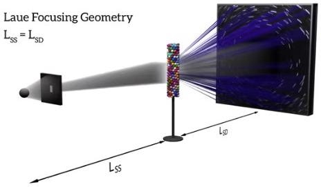
DCT projection images generate diverse diffraction patterns on the detector depending on their concentrating geometries. In Laue focusing mode, grains generate diffraction spots that are sharp lines on the scintillator-coupled objective-based detector.
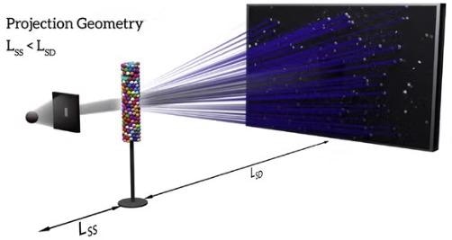
Projection geometry utilizes the flat panel detector placed in the defocused or magnifying spot to gather diffraction spots that look like protruding shape profiles of the equivalent diffracting grains.
The LabDCT Pro module on the ZEISS Xradia 620 Versa allows grain mapping on both the steadfast 4X DCT objective and on the optional flat panel detector offering the flexibility to select between high resolution or high throughput large area grain mapping while ZEISS Xradia CrystalCT uses its dedicated flat panel detector in projection mode.

LabDCT Pro: Laue focusing geometry with the 4X DCT objective. Image Credit: Carl Zeiss Microscopy GmbH

LabDCT Pro or CrystalCT: Projection or Laue focusing geometry onto a flat panel detector. Image Credit: Carl Zeiss Microscopy GmbH